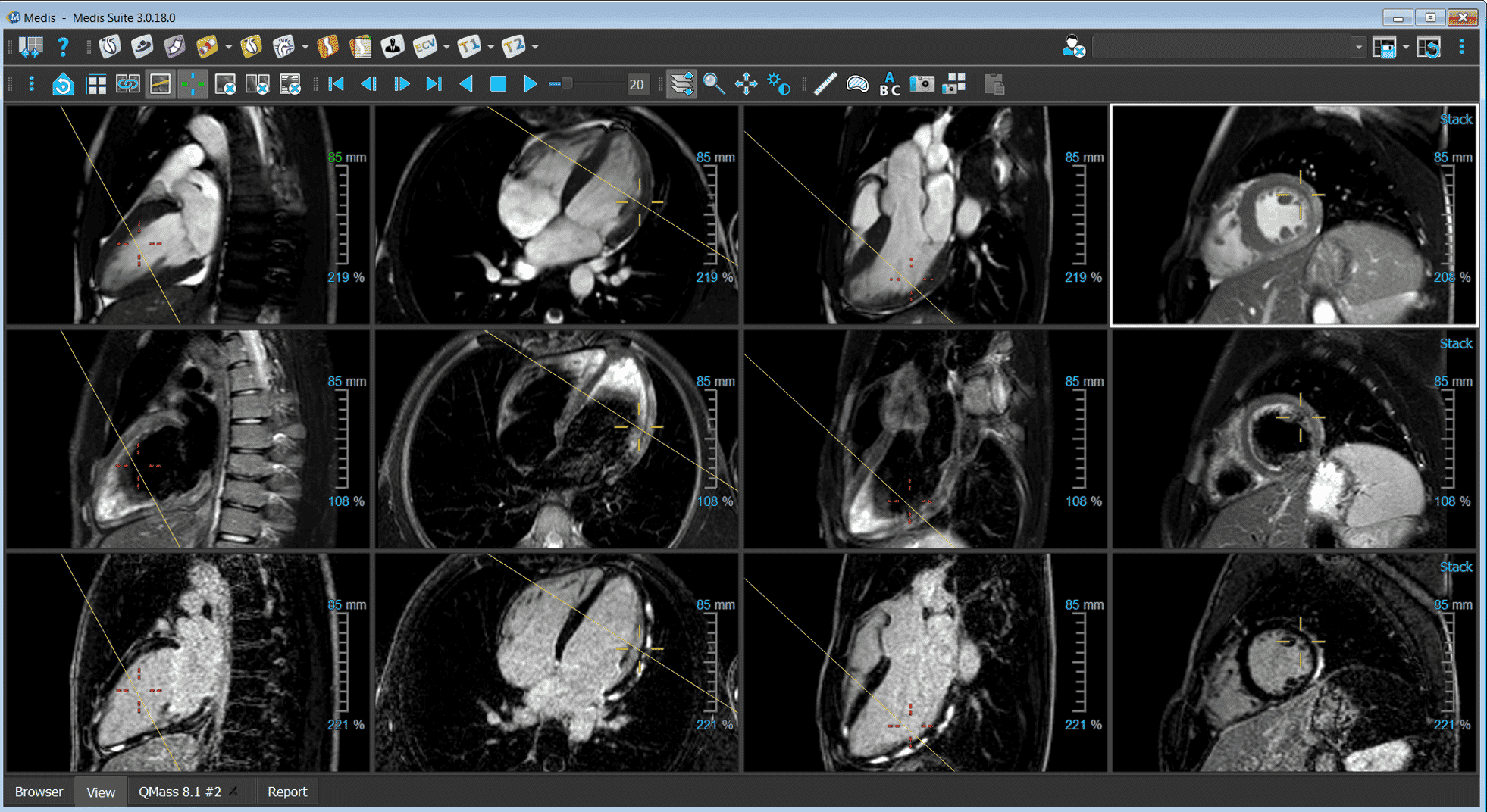
The first impression of the heart and its state is based on the anatomy over the cardiac cycle. With the 2D and 3D viewer you can see if there are congenital abnormalities, irregularities in the heartbeat, thickenings in the myocardium or check the quality of the MR scan.
DICOM Viewer
Get a first glance of the patient’s condition by viewing the MR series.
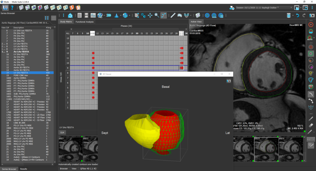
3D Viewer
Get a 3D visualization and/or reformat the 3D in a 2D series.
The cardiac systolic functions show you the current state of the heart, all based on the volumetrics. Additional deeper insights in the heart’s function can be gathered via the (global) deformation of the heart, via (regional) inward wall displacement, and intraventricular pressure gradients of the left ventricle.
LV/RV Function
Review the left and right ventricular function based on AI contours.
LV Strain
Assess left ventricle’s deformation with the global and regional strain based on feature tracking.
RV Strain
Assess right ventricle’s deformation with the global and regional strain based on feature tracking.
Atrial Strain
Assess the left and right atrium’s deformation with the global and regional strain values.
Inward Displacement
New parameter to assess the regional ventricular function based on feature tracking.
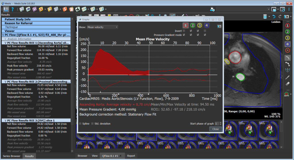
Evaluation of the blood flow in the greater vessels provides insights into the patient’s condition. Both with 2D flow and 4D flow blood flow volumes and velocities can be measured. Especially with 4D flow also the blood flow direction and velocity can be visualized to better interpret the results.
2D Flow
Assess the blood flow in the greater vessels on phase-contrast images.
4D Flow
Assess the blood flow in 3D fashion during the cardiac cycle.
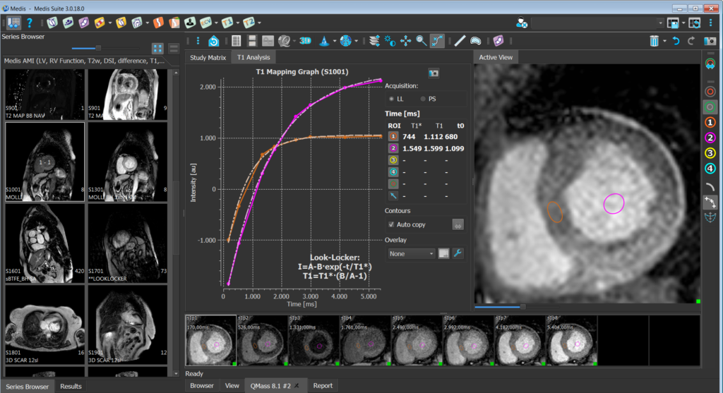
To diagnose the type of disease that is causing the reduced cardiac function different analysis have been developed, together they provide the entire picture. With LGE imaging scar can be detected, with T2weighted the edema in the heart, and with stress imaging the perfusion. But also, the mapping sequences help to differentiate between cardiac diseases.
T1 Analysis
Quantify T1 relaxation times in any given region of interest in the myocardium.
T2/T2* Analysis
Quantify either the T2 or T2* relaxation time in any region of the myocardium.
Delayed Signal Intensity (DSI)
Visualize and quantify the amount of damaged tissue based on the delayed signal intensity.
Time Signal Intensity (TSI)
Visualize and quantify the perfusion defect in rest and stress.
T1 Analysis
Research
Quantify T1 relaxation times in any region of interest or in the 16-segement model of the myocardium.
T2/T2* Analysis
Research
Quantify T2 or T2* relaxation times in any region of interest or in the 16-segement model of the myocardium.
Extracellular Volume
Research
Measure the extracellular volume based on T1 pre- and post-contrast scans.
Medis Suite MR stands as a comprehensive solution for cardiac MRI post-processing, crucial in both clinical and research settings. This platform excels in evaluating heart structures and functions with vendor-independent, advanced algorithms. It has powered global cardiologists and radiologists for over 25 years, contributing to 1500+ scientific cardiac MRI papers.
Medis suite MR includes innovative features like automated deep learning contour detection, flow analysis, and feature-tracking. Notably, MR Strain complements Echo and CT Strain, offering a multimodal approach for cardiac deformation imaging. It provides unique parameters for Inward Displacement and Hemodynamic Forces analysis. Medis Suite MR enables quantitative assessments of heart function, strain, perfusion, viability, mapping, and flow in 2D & 4D, integrating seamlessly into healthcare IT systems and DICOM networks.
Proven Accuracy
Powerful AI
Innovative
Robust Platform
What is new in the latest version of Medis Suite MR?
In-depth assessments of Left Atrium (LA) and Right Atrium (RA) with volumetrics, ejection fraction (EF), and strain analysis.
Advanced deep learning contouring covering also RV, LA, and RA for precise strain analysis.
Cutting-edge speed in cardiac volumetric and deformation analysis.
Applications of new features
LA Strain for systemic disease, cardiomyopathy, ischemic heart disease, atrial fibrillation, congenital heart disease, and HFpEF.
RA Strain for pulmonary arterial hypertension, ischemic heart disease, cardiomyopathies, and HFpEF.
Unique values tailored to your practice
Enhanced automation in QStrain, eliminating manual complexities with AI-powered contouring.
A new benchmark in diagnostic reporting, presenting critical information concisely for informed decision-making.
Simplification of complex hemodynamic forces (HDF) data into a clear visual narrative to review intra-ventricular pressure gradients (IVPGs) in the Research Edition
“Medis Suite MR has increased our ease of use, saved us a lot of time, and consequently increased the quality of clinical reporting”
“Medis Suite MR is automated, but also gives the flexibility to interact”
Prof. Atilla Toth
Interventional cardiologist / Cardiologist
“Medis Suite MR is our primary solution for reading CMR studies. Medis provides reliable and easy to use tools. We review cases with the team each day and find the viewer extremely valuable for these review sessions.”
Dr. David M. Leistner
Interventional cardiologist / Cardiologist
“Medis Suite MR improved our CMR post-processing workflow in terms of time and accuracy.”
Dr. Dietrich Beitzke
Interventional cardiologist / Cardiologist
‘The Medis machine learning with AutoQ contours is absolutely fantastic, a real game changer and a huge time-saver.’
Dr. Russell Bull
Interventional cardiologist / Cardiologist
“I especially like how visual and straightforward it is to select, add and edit planes for measurement. You can get good results fast without really noticing how much data there can be in the background.’”
Juha Peltonen
Medical Physicist
“We are using Medis Suite MR for research trials and our daily clinical work. In addition, the easy handling and proven accuracy of the measurements make the software excellently suited for our MRI Core Lab Berlin.”
Prof. Dr. Sebastian Kelle
Interventional cardiologist / Cardiologist
“Medis Suite MR is our primary solution for reading CMR studies. Medis provides reliable and easy to use tools. We review cases with the team each day and find the viewer extremely valuable for these review sessions.”
Dr. Raymond Kwong
Interventional cardiologist / Cardiologist
Request a demo of Medis Suite MR
You are only a click away from having your demo with an experienced Medis Suite MR product specialist.