Pressure

Pressure gradients in the ventricle play an important role in the ventricular filling. Currently, to measure these intraventricular pressure gradients (IVPG), a difficult and lengthy procedure is needed. With the Medis solution Hemodynamic Forces (HDF) this is no longer the case, as only non-invasive ultrasound B-Mode or MRI cine images are necessary. With this innovative novel method, Medis is pushing further the new frontier on cardiac deformation analysis and function assessment.

Pressure Gradients

Cardiac function relates to creating and sustaining blood motion. This is achieved through a sequence of contraction-relaxation in the myocardial muscle which creates pressure gradients across the ventricle allowing blood flow.

During the cardiac cycle, intra-ventricular pressure gradients are mostly directed from LV base to LV apex during diastole (except for late diastole), and from LV apex to LV base during systoleThe analysis of intracardiac hemodynamic force (HDF), which corresponds to the global value of Intraventricular Pressure Gradients integrated over the ventricular volume, offers a rigorous method to explore IVPGs and blood flow within LV [1]. These IVPGs play an important role in ventricular filling in the normal heart and may be abolished by systolic or diastolic dysfunction [2]. As flow-mediated forces act on tissue, they influence the ventricular remodeling.

Figure 1: The evolution of cardiac function analysis [3]
Figure 2: graphical representation of the pressure and flow distribution during the cardiac cycle

Medis can analyze the hemodynamic forces (HDF) non-invasively, where they were historically invasively measured with a catheter [2]. Even though more recently, this could be done by flow imaging, by echocardiography (color Doppler M-mode and Echo-PIV) or by 4D flow MRI. Both these techniques have disadvantages: for Echo-PIV injection of contrast agent is required and it is not a common clinical practice, while for the 4D flow MRI the lengthy scanning time, the low space and time resolution are limitations [3]. With Medis ultrasound and MRI solutions, the new Hemodynamic Forces module overcomes these limitations as a 3D output can be generated by the regular 3 long axis views. A dedicated mechanical model based on the LV endocardial contour with either speckle- or feature-tracking is applied to generate the HDF results [1]. 

Book your demo of Hemodynamics Forces

Pathologies

Myocardial Infarction

In patients with ST-segment elevation myocardial infarction (STEMI) it is difficult to identify adverse LV remodeling (aLVr) after reperfusion. During follow-up after 4 months, it has been shown that a higher diastolic HDF was associated with aLVr in STEMI patients [4]. This shows that HDF can be used as a predictor of post-infarction aLVr.

Figure 3: HDF distribution at follow-up of on the left a patients without adverse remodeling and right a patient with adverse remodeling [5]

Heart failure

Diagnosis

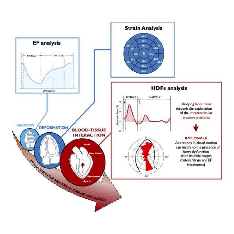
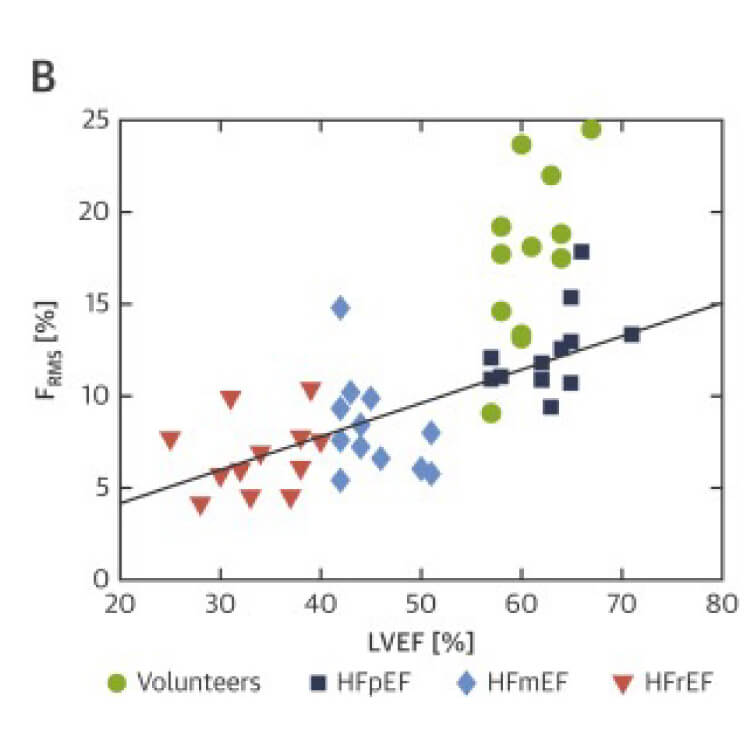
In case of heart failure with either mid-range or reduced ejection fraction (resp HFmEF and HFrEF) values of strain and ejection fraction can help with the diagnosis. However, those values can’t distinguish healthy volunteers and patients with heart failure with preserved ejection fraction (HFpEF) making early diagnosis challenging. The new HDF parameter shows promising results with a study by Lapinskas et al that indicates HDF may significantly improve the detection of early systolic dysfunction [5].

Treatment

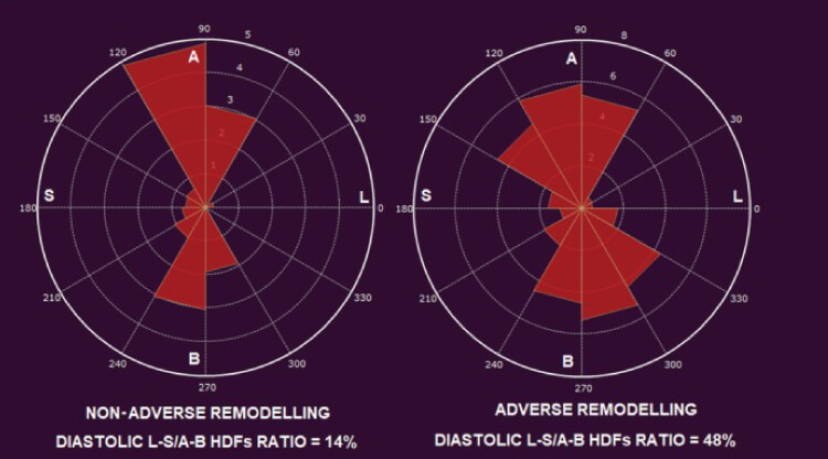
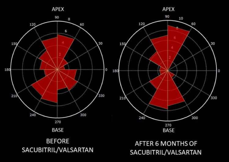
In case a patient is diagnosed with heart failure and medication therapy is prescribed, a follow-up on the evaluation of the cardiac remodeling is needed. It has been shown that for patients with heart failure with reduced ejection fraction (HFrEF) the HDF can be used as follow-up methodology. Patients treated with sacubitril/valsartan therapy showed re-alignment of the HDF after 6 months, indicating reverse remodeling of the LV [6].

For symptomatic heart failure patients without response on medical therapy, cardiac resynchronization therapy (CRT) is an established strategy. However, despite efforts to identify the appropriate candidates for CRT the response of the therapy is heterogeneous. Research shows that pacing-induced realignment of HDFs is associated with CRT efficacy at follow-up which might help with the patient selection [7] 

Figure 2: Bull's eye plots of speckle tracking longitudinal strain patterns in patients with amyloidosis [6]
Figure 5:Changes in HDF distribution before and after treatment of a HFpEF patient [4]

(non-)Hodgkin Lymphoma

A side effect of lymphoma cancer treatment can be cardiotoxicity, resulting in cardiovascular disease. Therefore, markers are needed to identify this risk or onset during treatment. HDF was investigated as a potential marker, and it turns out that the lateral-to-septal hemodynamic force was significantly lower in lymphoma survivors than in healthy controls [8]. This shows HDF might be a diagnostic indicator.

Measuring Hemodynamic Forces using Medis

The concept of IVPG has been there for a long time but the easiness to measure has been lacking. Medis saw the need to develop a simple and innovative method: the Hemodynamic ForcesResearch module based on speckle/feature tracking and valvular distances. With that your daily echo or MRI scanning routine is enough, and you no longer need additional scans or operating invasively. 

Medis Hemodynamic Forces solution derives the IVPGs in the left ventricle from “bread and butter” clinical images. It is available for our MR and Ultrasound solutions as a research add-on, enhancing insights into your patients’ cases. 

Jacqueline L Vos, Tim Leiner, Arie P J van Dijk, Gianni Pedrizzetti, Fawaz Alenezi, Laura Rodwell, Constantijn T P M van der Wegen, Marco C Post, Mieke M P Driessen, Robin Nijveldt, Cardiovascular magnetic resonance-derived left ventricular intraventricular pressure gradients among patients with precapillary pulmonary hypertension, European Heart Journal – Cardiovascular Imaging, 2022;, jeab294, https://doi.org/10.1093/ehjci/jeab294

Benefits of measuring pressure using Medis

Feature/Speckle Tracking

Medis’ tracking algorithms are developed by the Founders of strain. Both the speckle and feature tracking algorithms are robust, vendor-independent, and proven algorithms. Those algorithms have been optimized for each specific modality and have been used in over 1700 scientific papers.

Regular tissue imaging

To measure the IVPGs only regular long axis cine images are needed. Based on the feature tracking and valve dimensions in those images the hemodynamic forces can be calculated. Therefore, no additional time is needed during scanning or during the analysis as it reuses the tracking done for strain analysis.

Multi-modality

Hemodynamic Forces can be measured on Ultrasound, and MRI images with Medis. This enables comparing data from one modality to another, as for both modalities the same biomechanical modelling using endocardial tracking results as input for the hemodynamic forces. This Medis multimodality solution ensures that HDF can be achieved for each patient.

Using Hemodynamic Forces across modalities

Medis Suite MR [4]

Medis Suite Ultrasound

Robin Nijveldt - Dr - Cardiology - Medis Testimonial

Dr. Robin Nijveldt

Professor of Cardiovascular Imaging and Cardiologist at Radboud University Medical Center, Nijmegen, the Netherlands

1-on-1 interviews with our users

“At the Radboud UMC, we aim through research to better understand the underlying disease causes which can improve diagnosis or treatment in the end. With the HDF module of Medis, we have looked at patients with precapillary pulmonary hypertension to see if this parameter can help in the detection of subtle changes in the LV mechanics, which seems to be the case. This result makes us continue to further research the value of HDF.”

Robin Nijveldt is a Professor of Cardiovascular Imaging and Cardiologist at the Radboud University Medical Center (Nijmegen, the Netherlands), as well as connected to the Radboud Institute for Health Sciences. He works in a high-volume site, at the department of Cardiology focusing on the Vascular damage research theme, with a special interest in Cardiovascular Magnetic Resonance Imaging, CT and echocardiography, and in close collaboration with the department of Radiology.

He is interested in new imaging techniques and innovative strategies on today’s scientific health challenges with the aim to improve patient outcome in a personalized approach.

How long have you been using Medis Suite MR and what aspects attracted you to this product?

I have worked with Medis for 20 years now and have been using Medis Suite MR both for clinical purposes (LV volumes, Ejection fraction, Strain and Flow and 4Dflow for congenital patients) and research (stress perfusion quantification). I have seen Medis grow throughout the years and stayed in close contact with the team to share ideas and suggestions. I feel that Medis actively uses my input to improve the products and overall customer experience.

What are your thoughts on the latest HDF for Medis Suite MR?

At the Radboud UMC we aim through research to better understand the underlying disease causes which can improve diagnosis or treatment in the end. With the HDF module of Medis we have looked at patients with precapillary pulmonary hypertension to see if this parameter can help in detection subtle changes in the LV mechanics, which seems to be the case. This result makes us continue to further research the value of HDF.

What made Medis (Suite MR) stand out from other options in the market?

When you select a software solution/provider, it is highly likely that you are getting used to the way it works, you get trained on the particular features, functionality and workflow which makes it harder to switch to a different one. However, for me Medis Suite MR is quite intuitive that anyone could easily adapt.

Unleash the full power of heart function analysis

With all other parameters of Medis Suite MR, CT and Ultrasound

Measuring volume with Artificial Intelligence

Measuring shape deformation with strain

Measuring Motion with Inward Displacement

Book your Demo of Hemodynamics Forces

We are happy to give you an introduction to HDF, customized to your needs. We can showcase the research opportunities tailored to your hospital.  
Please fill out the form and Medis will shortly reach out to you.

    Name *

    Email *

    Country *

    * = Required