Book your Medis Demo
Do you want to learn more about Medis products? Contact us and we can set up a demo.
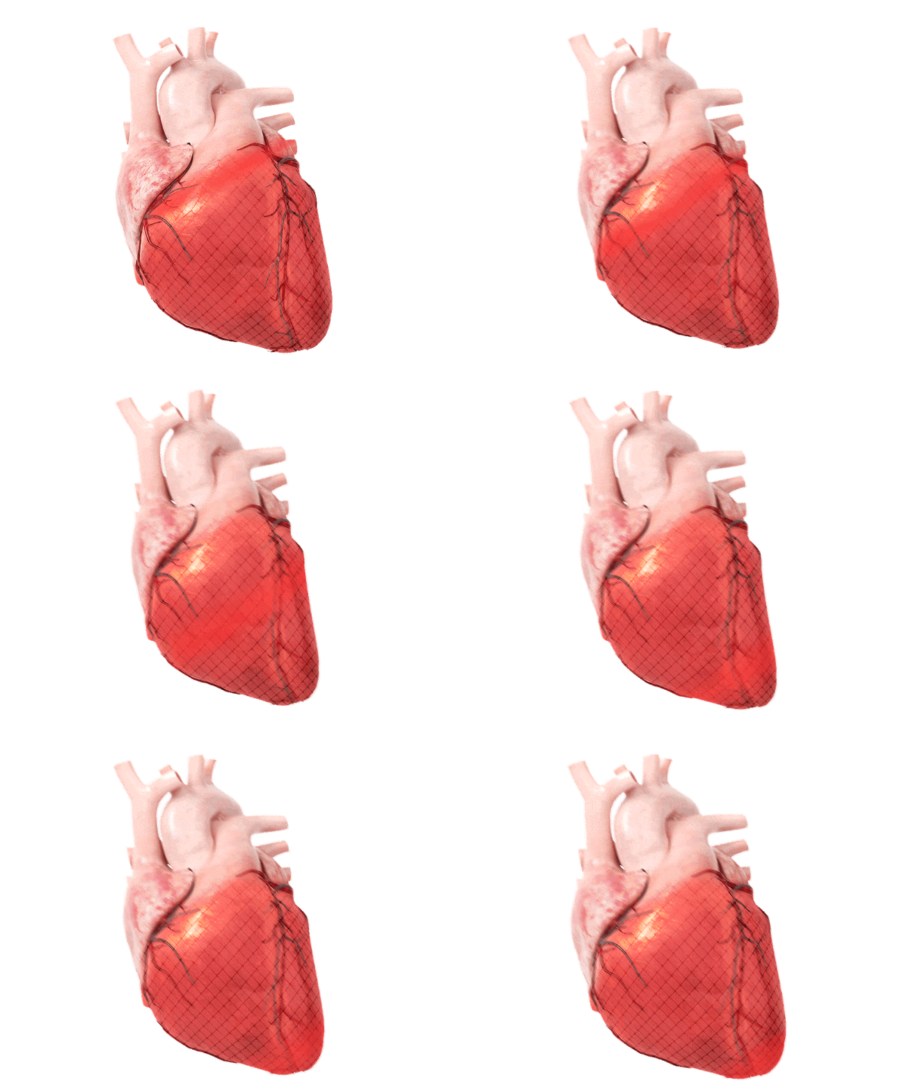
Cardiac volumetrics are the cornerstone in detecting cardiac function. It is measured in all cardiac patients which enhances the need for a quick and reliable solution. Medis Medical Imaging has empowered the MR function analysis with Deep Learning AI technology to simplify and speed up your daily analysis.

Nowadays, cardiac volumetric function analysis forms the foundation to assess and manage cardiac dysfunction [1]. MR imaging is the golden standard to measure the function analysis, volumes, and Ejection Fraction (EF), non-invasively. Normal values for each of the volumetric metrics have been defined to be able to establish dysfunction. For the EDV and ESV, this is gender and age-dependent, as supported by different papers such as Maceira et al [2], Sarikouch et al [3], and Peterson et al [4]. For the EF, a percentage of roughly 52% or higher is seen as normal and below 52% as impaired, although the cut-off value slightly differs per guideline [5].

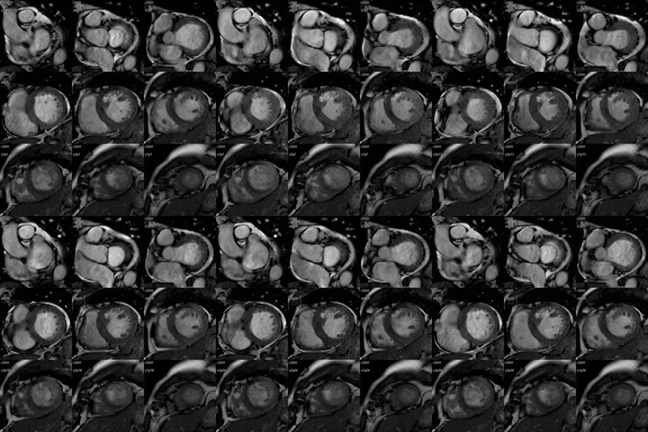
Cardiac Short Axis Series
For almost 30 years, Medis technology has helped advance the role of cardiac function analysis in clinical practice significantly. We are aware of the importance of accuracy and reproducibility when measuring volumetrics. Our Medis Suite MR solution is therefore empowered with Deep Learning AI technology to simplify and speed up your daily used function analysis.
Medis Function Analysis Explained by our Product Manager
Getting the endo and epicardial borders can be a time-consuming and repetitive task. With Medis 80% of our customer cases require no manual corrections making follow-up data better to interpret. This is all due to Medis AI Deep learning algorithm that has been trained on almost 1000 datasets in various cases across different vendors and pathologies.
Medis has incorporated different automation steps to shorten the analysis time. Such as automatic short axis series identification, automatic recognition of the DL contours (ED and ES phase), as well as calculation of the EDV, ESV, EF and all other volumetric values without manual interaction, after loading of series. All results are automatically synced and presented in the clinical report.

Do you want to learn more about Medis products? Contact us and we can set up a demo.
Medis Medical Imaging Systems BV
Schuttersveld 9, 2316 XG Leiden
The Netherlands
©2025 Medis Medical Imaging Systems B.V. All rights reserved.