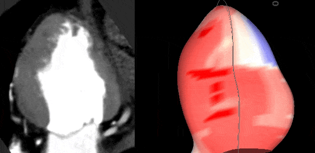
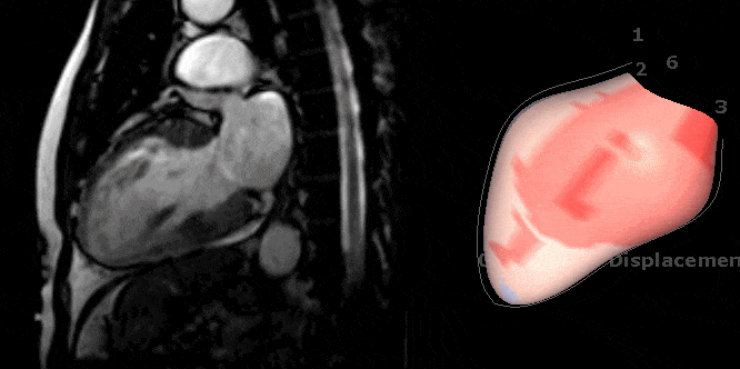
Multi-modality
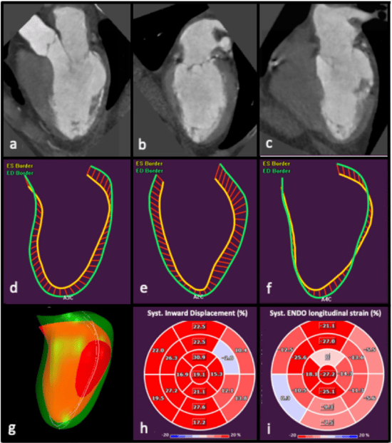
Inward Displacement can be measured on Ultrasound, MRI, and CT images with Medis. This enables comparing data from one modality to another, as for all modalities the inward displacement is based on the same algorithm., Medis multimodality solution, ensures that very likely you will have inward displacement data for each patient, depending on at least one available modality


"QStrain is a quick and easy solution, but I've found the bull's eye visualization not intuitive enough in a lot of cases. On the other hand Inward Displacement module with it's simple mechanical concepts reflects wall motion abnormalities better in my opinion. It can be especially helpful for those who are less experienced or experts who have to supervise multiple case."
Dr. Atilla Tóth Radiologist at Gottsegen György Hungarian Institute of Cardiology & Semmelweis University