Medis Suite MR

Cardiac diagnostics made simple.

Medis Suite MR is our comprehensive, time-saving and validated solution for cardiac MRI post-processing.

Anatomy

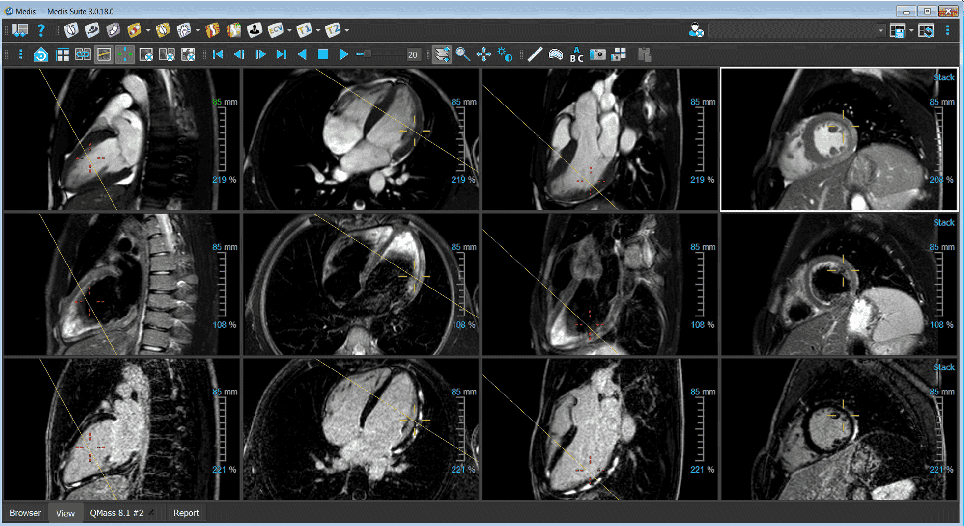
Explore the sophisticated anatomy of the heart throughout the cardiac cycle. Utilize our 2D and 3D viewers to identify congenital abnormalities, irregular heart rhythms, myocardial thickening, and assess the quality of MR scans, providing critical insights into cardiac health.

Quickly assess patient’s condition with MR series viewing.
Visualize and reformat 3D data for comprehensive analysis.
Cardiac MRI Medis Suite MR viewer

Cardiac function

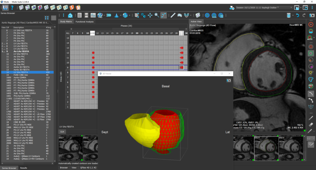
Gain valuable insights into the heart’s function through comprehensive assessment of its systolic functions, based on volumetric measurements. Dive deeper into cardiac function by analyzing global deformation, regional wall displacement, and intraventricular pressure gradients of the left ventricle, enabling a thorough evaluation of cardiac health.

Evaluate left and right ventricular function effortlessly with AI-driven contours.
Evaluate left ventricular deformation globally and regionally using feature tracking.
Assess right ventricular deformation globally and regionally with feature tracking.
Evaluate left and right atrial deformation globally and regionally using feature tracking.
New parameter for assessing regional ventricular function through feature tracking.
Obtain Intraventricular Pressure Gradients via Hemodynamic Forces based on feature tracking.
Medis Suite MR Clinical Applications LV/RV Function

Flow Analysis

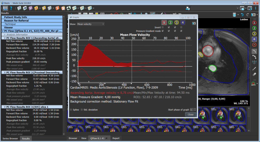
Evaluate blood flow in greater vessels to better understand the patient’s cardiovascular condition. Our 2D and 4D flow analyses allow precise measurement of blood volumes and velocities, while 4D flow visualization enables visualization of flow direction and velocity, enhancing interpretation of results and helping you in diagnosis.

Evaluate blood flow in greater vessels using phase-contrast images.
Analyze blood flow throughout the cardiac cycle in 3D.

Tissue Characterization

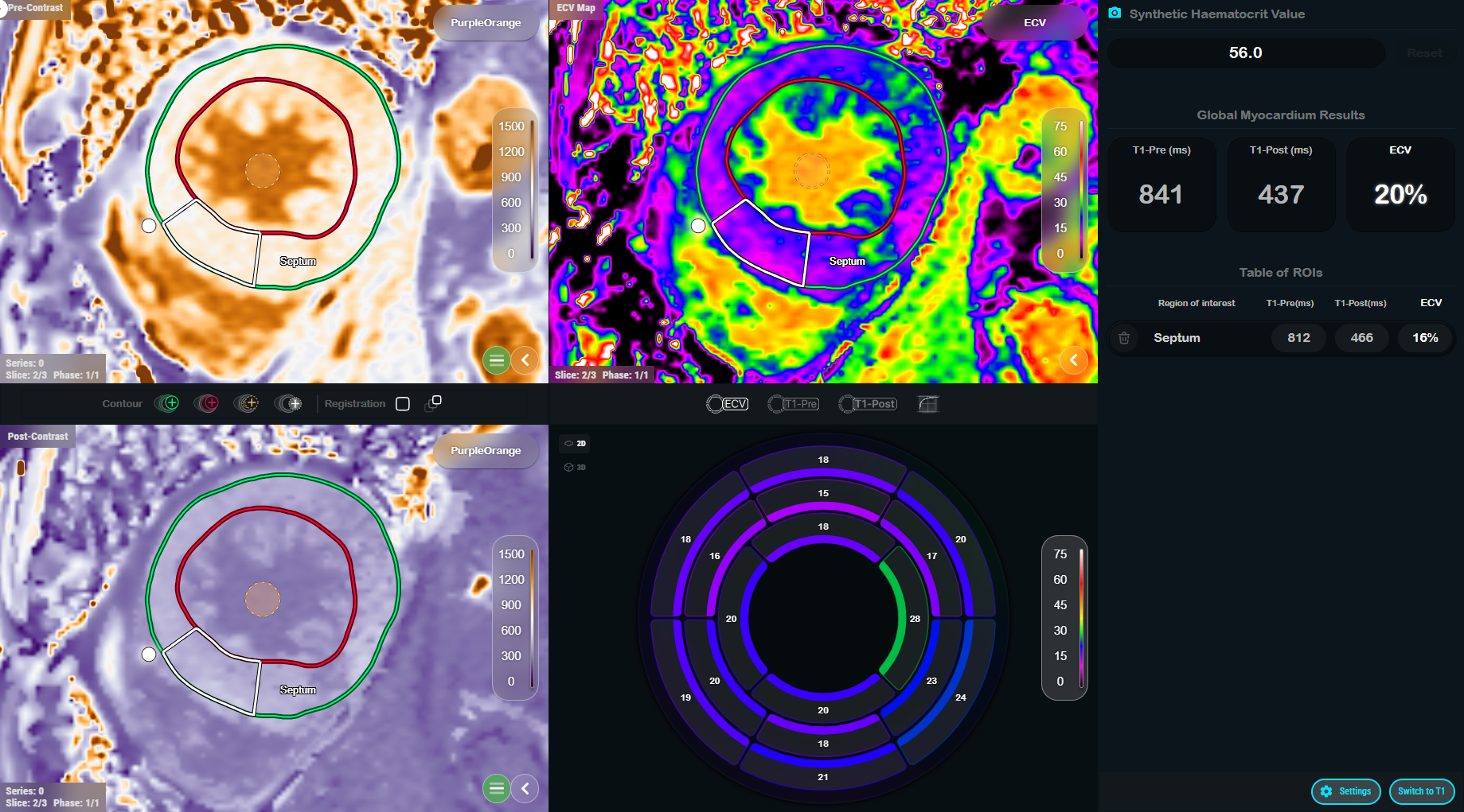
Diagnose underlying cardiac diseases contributing to reduced cardiac function through advanced tissue characterization analyses. Detect scar tissue with LGE imaging, identify edema with T2-weighted imaging, and assess perfusion with stress imaging. Additionally, mapping sequences help differentiate between various cardiac pathologies, providing a comprehensive diagnostic approach.

– Fully automated T1 & ECV Mapping

– Automatic motion correction and co-registration

– Synthetic hematocrit enabled ECV

1The ECV functionality is for research use only in CE countries.

Quantify either the T2 or T2* relaxation times in any region of the myocardium.

Visualize and quantify damaged tissue using delayed signal intensity.

Visualize and quantify the perfusion defects at rest and stress.

Quantify T2 or T2* relaxation times in any region of interest or in the 16-segment model of the myocardium.

Medis Suite MR

Medis Suite MR is an extensive, validated and time-saving cardiac MRI post-processing solution, ideal for both clinical and research settings. Trusted by cardiologists and radiologists worldwide for over 30 years, and featured in more than 1500 publications, Medis Suite MR excels in evaluating heart structures and functions with advanced algorithms powered by AI. Vendor-independent, Medis Suite MR enables quantitative assessments of heart function, strain, perfusion, viability, mapping, and flow in 2D & 4D, all seamlessly integrated into healthcare IT systems and DICOM networks.

Proven Accuracy

AI-driven Efficiency

Continuous Innovation

Integrated Diagnostic Workflow

What is new in the latest version of Medis Suite MR?


Designed to make advanced tissue characterization faster, simpler, and more accessible.


Built on decades of cardiovascular MR expertise and shaped by customer feedback, T1 & ECV1 Mapping is the first Medis Suite MR application redesigned on our next-generation software architecture. 

It works seamlessly alongside existing Medis Suite MR applications while introducing a modern user experience and establishing the foundation for future innovations across the suite.

Release highlights

– Efficient ECV1 calculation without blood sampling through synthetic hematocrit computation

– Streamlined workflow for efficient tissue characterization

– Intelligent automation with transparent clinical review

– Advanced tissue quantification with customizable, easy-to-interpret visual outputs, supporting greater confidence in image interpretation

1The ECV functionality is for research use only in CE countries.

“Medis Suite MR has increased our ease of use, saved us a lot of time, and consequently increased the quality of clinical reporting.”

Dr. Robin Nijveldt

Cardiologist

“Inward displacement is a great new tool that will allow to quantify wall motion abnormalities in an objective way”

Dr. Erasmo de la Peña-Almaguer

Cardiologist

“Medis Suite MR is automated, but also gives the flexibility to interact”

Prof. Atilla Toth

Radiologist

“Medis Suite MR improved our CMR post-processing workflow in terms of time and accuracy.”

Dr. Dietrich Beitzke

Radiologist

“Medis Suite MR is our primary solution for reading CMR studies. Medis provides reliable and easy to use tools. We review cases with the team each day and find the viewer extremely valuable for these review sessions.”

Dr. Raymond Kwong

Radiologist

Brigham Health logo

Request a free demo of Medis Suite MR

You are only 1 click away from having your demo with an experienced Medis Suite MR product specialist.