
The Metrics of the Heart
Overview
The Metrics of the Heart: measuring cardiac function
Key takeaways
- Measuring the shape deformation or myocardial strain of the heart is becoming an integral part of studying cardiac function, providing global longitudinal strain (GLS), global circumferential strain (GCS) and global radial strain (GRS).
- What is cardiac function? Cardiac function refers to the ability of the heart to efficiently pump blood throughout the body, which can be measured through a variety of metrics including myocardial strain, inward displacement, and hemodynamic forces, providing valuable information beyond commonly used volumetric measurements and aiding in the diagnosis, treatment, and follow-up for patients with cardiac disorders.
- Regional strain provides information on regional ventricular dysfunction, but the sensitivity of regional strain has been shown to be relatively low. Medis has created an actual left ventricular wall motion model to detect regional wall motion abnormalities, which allows for an objective quantitative assessment of regional cardiac function.
- The analysis of intracardiac hemodynamic force (HDF) or Intraventricular Pressure Gradients (IVPG) offers valuable information about the effectiveness of the cardiac function.
- Medis provides AI-based tools to measure these additional metrics – strain, inward displacement, and hemodynamic forces – to guide the diagnosis, treatment, and follow-up for patients.
- These additional metrics add valuable information to better understand changes in cardiac function, beyond the commonly used volumetric measurements, and are available for multiple modalities including MR, CT, and ultrasound.
For years now, the volumetrics have been the most commonly used clinical measurements on how the heart functions, for which Medis provides AI based tools. However, scientific evidence indicates that there is more than just the volume to measure the cardiac function.
The shape, the wall motion and the pressure gradients of the heart are all influencing the effectiveness of the cardiac function. At Medis we recognize the need to look beyond the volumetrics of the heart and equip you with the tools to determine changes in the cardiac function from all angles. Medis offers besides the regular ejection fraction, three additional ways to extent the cardiac function analysis in a multimodality approach.
- Strain
- Inward Displacement
- Hemodynamic Forces
Measuring these additional metrics adds valuable information to better guide the diagnosis, treatment and follow up for your patients. The way you used to establish cardiac function is going to change!
1. Measuring Shape Deformation
The cardiac shape deformation, myocardial strain, is becoming an integral part of studying cardiac function and is extensively used as a parameter to describe the function of the left ventricle. Strain measures the myocardial tissue shortening during systole in longitudinal, circumferential, and radial directions, providing global longitudinal strain (GLS), global circumferential strain (GCS) and global radial strain (GRS). In contrast to ejection fraction (EF), assessment of longitudinal systolic deformation, global longitudinal strain, is a more sensitive tool in detecting early systolic dysfunction since changes in the EF may occur in later stages of several cardiac diseases [1, 2].
Strain can be measured both on a global and a regional scale, the latter based on the AHA segment model. In addition, as the function of the heart is determined by all its chambers, strain metrics are of value for both left and right ventricles as for the atria.
Although strain, especially global longitudinal strain, is well known in the echocardiography area, the principles have extended to the fields of cardiac magnetic resonance imaging (CMR) and Multi-Slice Computed Tomography (MSCT).
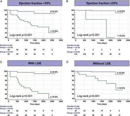
The value of global longitudinal strain keeps increasing by new evidence that is gathered through research highlighting the incremental prognostic value for the cardiac function in various cardiac disorders. For example, in dilated cardiomyopathy (DCM), both speckle tracking echocardiography and Feature Tracking (FT) CMR showed that depressed GLS can be used as a prognostic value to assess the response to therapy and predict the occurrence of major cardiovascular events [11, 12]. For FT-CMR, GLS was even found to be an independent indicator of survival in DCM patients beyond standard CMR values such as EF. As demonstrated in the image (Figure 1), DCM patients with preserved GLS exhibited excellent long-term outcomes despite the presence of severely impaired LV [13].

The Medis Strain solution is based on state-of-the-art speckle- and feature tracking- technology. This specific tracking-approach has been based on solid scientific principles and has demonstrated its accuracy and reproducibility within and across the different modalities. Its innovative technology has appeared in several hundreds of peer-reviewed studies and in some cases have become industry standard.
2. Measuring Wall Motion
Regional strain provides information on regional ventricular dysfunction by measuring the changes in parallel displacement along the left ventricular circumference. This provides insights in case of regional wall motion abnormalities. However, the sensitivity of regional strain has been shown to be relatively low. This is due to the fact, that small changes in the length of a subsegment along the perimeter of the left heart chamber are prone to large variabilities, depending on the definitions of these subsegments.
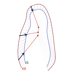
Over the years, various left ventricular wall motion models have been developed, the most well-known model is the centerline model [4]. This model is based on the changes in position of individual points along the LV perimeter from ED to ES and perpendicular to the centerline between the ED and ES contours. However, this method not based on a solid wall motion model.
The feature tracking technology provides much more information about the actual displacement of the individual positions along the left ventricular boundary. Based on that solid technology, Medis has created, together with our KOL’s, an actual left ventricular wall motion model to detect regional wall motion abnormalities which shows that points along the endocardial border are moving towards well-defined “centers of contraction”. Medis’ new Inward Displacement model allows for an objective quantitative assessment of regional cardiac function. Figure 3 shows a graphical representation of the basic principles of the Inward Displacement model.
Recently, a case report was published demonstrating the use of Inward Displacement to objectively confirm regional dysfunction in a patient with a myocardial infarct [5]. Assessing left ventricular remodeling and defining therapeutic strategies could be done based on the regional wall motion results.
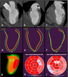
The Medis Inward Displacement solution can measure regional wall motion abnormalities in the left ventricle, and it is available for MR, CT, and Ultrasound. All to further enhance insights into your patients’ cardiac function. The Inward Displacement bull’s eye plot aims to describe the regional impairment most in line with visual observations.
3. Measuring Pressure Gradients
Cardiac function relates to creating and sustaining blood motion. This is achieved through a sequence of contraction-relaxation in the myocardial muscle which creates pressure gradients across the ventricle allowing blood flow (see figure 5).
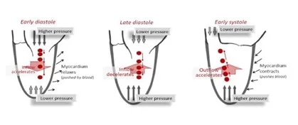
The analysis of intracardiac hemodynamic force (HDF), which corresponds with the global value of Intraventricular Pressure Gradients (IVPG) integrated over the ventricular volume, offers a rigorous method to explore IVPGs and blood flow within LV [6]. These IVPGs play an important role in ventricular filling in the normal heart and may be abolished by systolic or diastolic dysfunction [7]. As flow-mediated forces act on tissue, they influence the ventricular remodelling and thereby adding insights in the overall cardiac function.
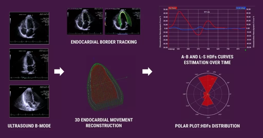
Historically the hemodynamic forces (HDF) were invasively measured with a catheter [7]. More recently, this could also be done by echocardiography (color Doppler M-mode and Echo-PIV) or by 4D flow MRI. Both these techniques have disadvantages: for Echo-PIV injection of contrast agent is required and it is not a common clinical practice, while for the 4D flow MRI the lengthy scanning time, the low space and time resolution are limitations [8]. With Medis’ ultrasound and MRI solutions, the new Hemodynamic Forces module overcomes these limitations as a 3D output can be generated by the regular 3 long axis views. A dedicated mechanical model based on the LV endocardial contours with either speckle- or feature-tracking is applied to generate the HDF results [6] [9].
This new module has been used already in different papers to show the added value in either diagnosis or treatment of the cardiac function. Recently a paper by Backhaus et al [10] has been published on hemodynamic forces in heart failure patients with preserved ejection fraction. This paper showed that the hemodynamic forces value was superior to LV global longitudinal strain in the prediction assessment for cardiovascular mortality and hospitalization in these patients.
Medis Hemodynamic Forces solution derives the IVPGs in the left ventricle from “bread and butter” clinical images. Therefore, you no longer need additional scans or operating invasively to enhance your cardiac function. HDF is available for our MR and Ultrasound solutions as a research add-on, enhancing insights into your patients’ cardiac function.
4. Benefits of using these additional values to measure cardiac function using Medis
Besides getting additional data from each of the above described enhanced measures for cardiac function and its benefit of being able to better understand the severity, location and status of the left ventricular dysfunction. All these modules are designed in such a way that the Medis software provides you additional benefits.
Multi-Modality
Medis global and regional strain, and Inward Displacement (wall motion) can be measured on MRI, CT and Ultrasound images. In addition, Medis’ Hemodynamic Forces can be measured on Ultrasound and MRI images. This enables comparing cardiac function data from one modality to another, as the same biomechanical modelling using endocardial tracking results are used as input for the measurements. This Medis multimodality solution ensures that you can have at least cardiac function insights from one modality for each patient.
Powered by AI
Getting the endo and epicardial borders for the cardiac function can be a time-consuming and repetitive task. With Medis deep learning contours, 80% of our customer cases require no manual corrections making follow-up data better to interpret and increasing reproducibility. This is the result of training the Medis AI Deep Learning algorithms for MR on almost 1000 datasets in various cases across different vendors and pathologies.
Automation
Medis has incorporated different automation steps to shorten the cardiac function analysis time and optimize the reproducibility. The main step in MR is the transfer of the automatic generated left ventricular contours (ED and ES phase) from the volumetric analysis. In addition, all results are automatically calculated and presented in the clinical report.
Speckle/Feature Tracking
Medis’ speckle and feature tracking algorithm are developed by the founders of strain. Both the tracking algorithms are proven, robust and vendor independent. Those algorithms have been optimized for each specific modality and overall have been used in over 1700 scientific papers.
5. Summary: measuring cardiac function
Medis recognizes the need to look beyond volumetrics and equip clinicians with tools to determine changes in cardiac function from all angles. For years, volumetrics has been the most commonly used clinical measurement of cardiac function. However, scientific evidence indicates that the shape, wall motion, and pressure gradients of the heart all influence the effectiveness of cardiac function. Medis offers three additional ways to extend cardiac function analysis in a multimodality approach: Strain, Inward Displacement, and Hemodynamic Forces.
Measuring shape deformation, or myocardial strain, is becoming an integral part of studying cardiac function. Strain measures the myocardial tissue shortening during systole in longitudinal, circumferential, and radial directions, providing global longitudinal strain (GLS), global circumferential strain (GCS), and global radial strain (GRS). Unlike ejection fraction, assessment of longitudinal systolic deformation, global longitudinal strain, is a more sensitive tool in detecting early systolic dysfunction. Strain metrics are of value for both left and right ventricles as for the atria. Medis Strain solution is based on state-of-the-art speckle- and feature-tracking technology. Its innovative technology has appeared in several hundreds of peer-reviewed studies and in some cases have become industry standard.
The Inward Displacement model is a solid wall motion model that provides much more information about the actual displacement of the individual positions along the left ventricular boundary. It allows for an objective quantitative assessment of regional cardiac function. Assessing left ventricular remodeling and defining therapeutic strategies could be done based on the regional wall motion results.
The analysis of intracardiac hemodynamic forces, which corresponds with the global value of Intraventricular Pressure Gradients (IVPG), is an innovative method to quantify the function of the heart. By using computational fluid dynamics, one can analyze the intraventricular pressure and blood flow over the complete cardiac cycle. This provides insight into the LV filling, pumping capacity, and flow distribution. The Medis Hemodynamic Forces tool is used to measure pressure gradients, providing a more comprehensive understanding of cardiac function.
Measuring these additional metrics adds valuable information to better guide the diagnosis, treatment, and follow-up for patients. With these additional tools, clinicians can now determine changes in cardiac function from all angles, going beyond volumetrics alone.
6. References
- https://www.ncbi.nlm.nih.gov/pmc/articles/PMC6209440/
- https://link.springer.com/article/10.1007/s10741-017-9621-8
- https://doi.org/10.1007/s10554-020-01883-9
- https://doi.org/10.1161/01.CIR.74.2.293
- https://doi.org/10.1007/s10554-021-02435-5
- https://doi.org/10.1161/JAHA.121.023417
- https://pubmed.ncbi.nlm.nih.gov/22114063/
- https://www.nature.com/articles/nrcardio.2014.75#citeas
- https://doi.org/10.1016/j.ijcard.2022.11.019
- https://doi.org/10.1016/j.ebiom.2022.104334
- https://www.sciencedirect.com/science/article/pii/S1936878X15008451?via%3Dihub
- https://www.ahajournals.org/doi/full/10.1161/CIRCIMAGING.109.910893
- https://academic.oup.com/ehjcimaging/article/16/3/307/2399809?
Share this article on: