
Medis Suite’s atrial strain analysis module boasts a robust algorithm that has been developed by key opinion leaders in the field. This algorithm provides state-of-the-art feature tracking technology to accurately track the myocardial from cardiac CT image series of the LV (that can be reformatted to LAX or SAX, RV in 4CH, LA in 2CH or 4Ch and RA in 4CH) enabling strain measurements. This approach ensures reliable and precise measurements, providing healthcare professionals with confidence in the accuracy of their diagnostic assessments.
Efficient Processing: Contour Re-usage
Medis Suite ventricular strain analysis is not limited to CT imaging alone. The software also enables strain analysis using the same post-processing method for MR or Ultrasound images. This versatility allows healthcare professionals to analyse ventricle strain from CT, MR and Ultrasound studies using a consistent approach. The ability to
compare CT and MR function studies enhances diagnostic confidence by allowing for comprehensive assessment and comparison of cardiac function across different imaging modalities.
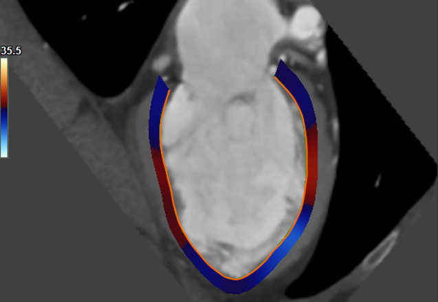
Medis Suite’s strain analysis module enables precise quantification of strain parameters. In both long-axis and short-axis orientations, the software quantifies strain parameters, including Global Longitudinal Strain (GLS), Global Circumferential Strain (GCS), and Global Radial Strain (GRS). Additionally, Medis Suite allows quantification of delta rotation, providing additional insights into cardiac mechanics. The software also facilitates the quantification of strain parameters, including strain, strain rate, and velocity, all are reported in the 16 segment American Heart Association (AHA) model. The ability to generate results for the endocardial, myocardial, and epicardial
wall further enhances the comprehensive assessment of LV strain.
Medis Suite CT generates detailed results from atrial strain analysis, allowing healthcare professionals to delve into the quantitative data. The software provides next to the clinical output an option to export detailed results to MS-Excel, facilitating further analysis and customization as needed. This export capability enhances data collecting, supporting research to get additional insights of the atria and its connection with entire cardiac function.
Medis Suite CT ensures simplified reporting by including key strain analysis results in its reports. This feature allows healthcare professionals to generate comprehensive and concise reports that highlight crucial atrial strain parameters. The inclusion of these key results in Medis Suite reports promotes efficient communication and facilitates a clear understanding of cardiac function.
You are only a few clicks away from having a demo with an experienced Medis Suite CT product specialist