
The LV/RV function analysis stands out as a powerful tool for accurately assessing the left ventricle (LV) and right ventricle (RV) function. This advanced functionality provides accurate and reproducible results.
The LV/RV function analysis feature within Medis Suite CT ensures precise and reliable measurements. By leveraging the software’s robust algorithms and validated methodologies, clinicians can trust in the accuracy of the obtained results, enabling them to make informed clinical decisions with confidence.
One of the key advantages of Medis Suite CT is its ability to facilitate a seamless comparison of results obtained from magnetic resonance (MR) and computed tomography (CT) images. By utilizing the same post-processing software, the same workflow and calculations are used to get the cardiac function, making the comparison more reliable.
Medis Suite CT’s 3D View application serves as a valuable reformatter allowing users to obtain multi-phase multi-slice series out of the cardiac CT stack. With this capability regular short and long axis cine images for the function analysis can be created.
Medis Suite CT further extends its capabilities by offering seamless integration with strain analysis based on CT feature tracking. Clinicians can effortlessly load the contours traced during the LV function analysis into the strain software. This integrated approach streamlines the workflow, saving valuable time and effort for cardiologists and radiologists.
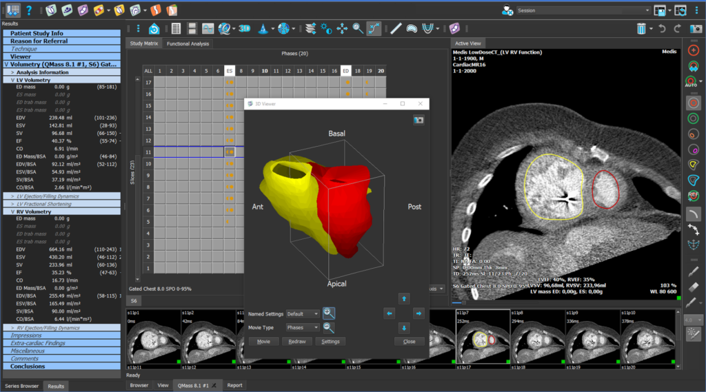
The LV/RV function analysis feature within Medis Suite CT offers an array of advanced functionalities to enhance cardiac assessment. It enables function analysis using Simpson’s method on short axis or transversal stack cine images for left and right ventricle, in addition also custom volumes can be added to get atrial volumes for example. Clinicians can quantify left ventricular volumes in the long axis using area-length and bi-plane volumetric analysis.
The software ensures automatic contour detection of LV endo- and epicardium, RV endocardium, and semi-automatic contour editing for precise measurements. The “LiveContour” algorithm allows for quick endocardial contour detection, further expediting the analysis process.
Medis Suite CT provides a comprehensive set of quantification parameters, including EDV, ESV, SV, %EF, CO, CI, indexed values, peak filling and ejection rates, and ejection rates, with various BSA calculation methods. The software also supports the calculation of z-scores for various normal ranges, enabling clinicians to assess LV/RV function in the context of age and sex specific reference values. Additionally, the software offers analysis of regional parameters such as wall motion, wall thickness, wall thickening, and wall thickness changes over time, providing a detailed assessment of cardiac performance.
You are only a few clicks away from having a demo with an experienced Medis Suite CT product specialist