
News
Medis Tips, Tricks & Shortcuts 3rd edition
02-02-20223
Welcome to the second edition of Medis Tips, Tricks & Shortcuts, a news section that addresses any questions of yours across all our product lines.
Twice a year we share a short survey in which we ask our customers to rate “how likely are you to recommend Medis” and provide us with general feedback.
We received many insightful comments that helps us keep improving in the right direction! In return, we would like to thank you by introducing this newsletter on a regular basis to ensure all your questions are addressed.
Did you miss the first two editions? No worries, you can find it here.
In this edition, we have summarized the next 5 questions that can be addressed immediately:
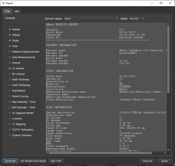
1. Is it possible to export results of QMass in an excel format?
Yes, this is possible. In QMass there is an advanced reporting system. You can find this via the shortcut F9 or via the 3 dots in QMass – View – Report. In the new pop-up screen, you can select which results you want to see with the checkboxes and select the desired level of detail with a dropdown menu.
After you have generated the results, you can save the entire file in either .txt or .csv format. If you only want to save a specific part, you can select this area and with a right mouse button click you can select the option ‘Copy text’ or ‘Copy CSV’.
2. Is there a manual or document for the RV function?
For all analyses in Medis Suite MR, detailed how-to instructions can be found in the Medis Suite MR user manual.
You can find the manual within the Medis Suite area. Go to the 3 dots , Help – User documents. A new tab will open where you can click on the user manual or a quick start manual in a specific language. There is also a function analysis movie as part of the MR explained, containing information on the RV function. Click here to watch it
3. Is it possible to measure the atrial volumes in the longer axis?
There are two approaches to measure the atrial volumes from the long axis within the Medis Suite MR application: The first approach is within the QMass application. This method is well described in the paper of Ma, Yanyan et al. The second approach is by performing an atrial strain analysis in QStrain, in which, based on the drawn contours, a volume curve is presented. This is also used in the paper of Chen, Xi et al.
4. When do I need to correct contours?
Focus on side branches, vessel overlaps and narrowing’s at the start/end points. The latter might be corrected automatically already by the software. Only edit a lesion when the contour is clearly incorrect. For vessel segments which are well filled with contract medium, the automatic contour detection is much better that the human eye. Do not zoom in; in the contour step, the vessel is automatically zoomed to view the contour optimally. In general, for reproducibility purposes, the less contour edits, the better.
5. Why should the offset location be in the middle of the analysis segment?
The offset location is used together with the start and end locations to calculate the 3D model. The software “knows” these locations are the exact same locations in both views. Therefore, if the offset location is the same as (or very close to) the start or end location, the software has only two instead of three locations to calculate with, making the 3D reconstruction less accurate.
If you want us to address a specific question in the next Tips, Tricks and Shortcuts please let us know here
Share this article on: