News

Medis Tips, Tricks & Shortcuts 2nd edition

13-12-2022

Welcome to the second edition of Medis Tips, Tricks & Shortcuts, a news section that addresses any questions of yours across all our product lines. 

Twice a year we share a short survey in which we ask our customers to rate “how likely are you to recommend Medis” and provide us with general feedback.  

​​​​​​We received many insightful comments that helps us keep improving in the right direction! In return, we would like to thank you by introducing this newsletter on a regular basis to ensure all your questions are addressed.
​​​​
Did you miss the first one? No worries, you can find it here

In this edition, we have summarized the next 5 questions that can be addressed immediately:

 1. Can Medis Suite export via structured reporting?  

Yes, all clinical results of MR and XA which are available in the report can be exported via DICOM Structured Reporting (DICOM SR). In the export section of the Medis Suite options, you can select the different export options. One of them is regarding the results to export them as DICOM Structured Report.  

Tips n tricks 2nd edition

2. Is it possible to measure LV thickness in each segment? 

Yes, it is possible to get the LV thickness for the AHA 16 segment model.  

How to: After you have done a function analysis based on the short axis you can use the reference point tool 
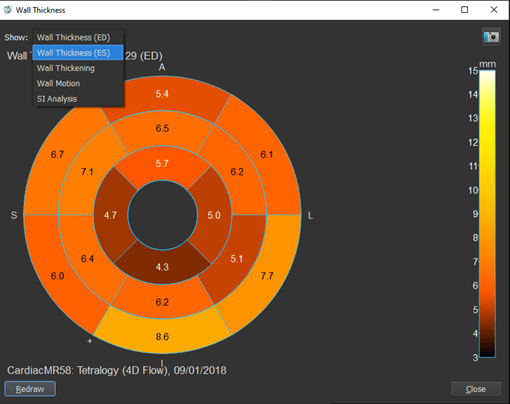
to locate the inferior intersection point between the LV and RV. Based on this the deviation in segments will be made based, also on the number of slices. Select this bull’s eye icon (with the Q of quantification) and you will see by default the wall motion 16 segment results. By pressing the dropdown menu in the pop-up screen, you can select the LV Thickness at ED or at ES.

Tips n tricks 2nd edition

Depending on your aim with the numbers you can get them via a snapshot in your clinical report via XML or JSON output in your reporting system, or via the advanced reporting tool in QMass into a text or CSV file.  

3. What can Medis detect with AI contour detection? Also, is it possible to get the LV strain without having to trace the contours? 

Medis Suite MR contains two segmentation algorithms supported by deep learning. One for the short axis, containing both LV and RV contours and one for the long axis containing LV contours. As a result, these functional analyses can be performed  with minimal editing; the feedback from our users is that this has improved their workflow significantly, as they don’t need to edit in 8/10 cases. 

In addition, we can forward the (edited) contours from the function analysis directly towards the strain solution;  as a result, you do not need to manually trace the contours for your strain analysis.  

4. Is it possible to enable the query retrieve at the series level? Our exams can surpass 10k images, we do not need to retrieve them all.​​

Within Medis Suite we cannot query and retrieve on series level, only the entire study can be retrieved. We have noted your request and hope that in the meanwhile the following work around will make sure your system will not occupy all the disk space. Instead of selecting the series before the retrieve, you can select the series after retrieval to be removed. 

How to: When the entire series is retrieved, click on all the series in the series info you don’t need. By pressing the Ctrl button, you can select multiple series at the same time or with the Ctrl + Shift button pressed select a range of series simultaneously. Once you have selected all the series you want to delete, click with your right mouse button, and select the option delete. After you have accepted it in the pop-up, all selected series will be deleted and no longer use disk space. ​​​​​​

5. When do I need to correct contours ?

Focus on side branches, vessel overlaps and start/end point narrowings. The latter might be corrected automatically already. Only edit a lesion when it is clearly wrong. With good contrast, the automatic contour detection is much better than the human eye. Do not zoom in; in the contour step, the vessel is automatically zoomed to view optimally. In general, for reproducibility, the less contour edits, the better.


If you want us to address a specific question in the next Tips, Tricks and Shortcuts please let us know here​​

Share this article on:

LinkedIn
Twitter

Sign up for our newsletter

Subscribe now to find out more about our solutions, latest publications and future releases.

By providing your information and continuing, you agree to Medis’ Terms of Use and Privacy Policy *