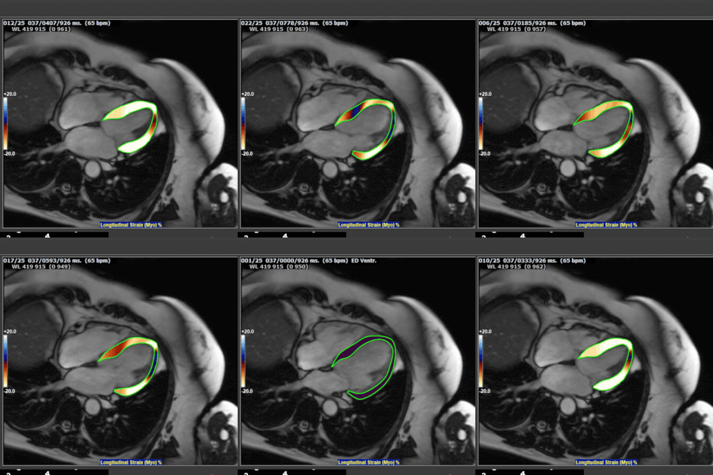
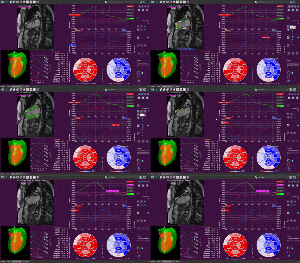
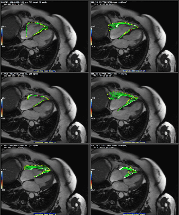
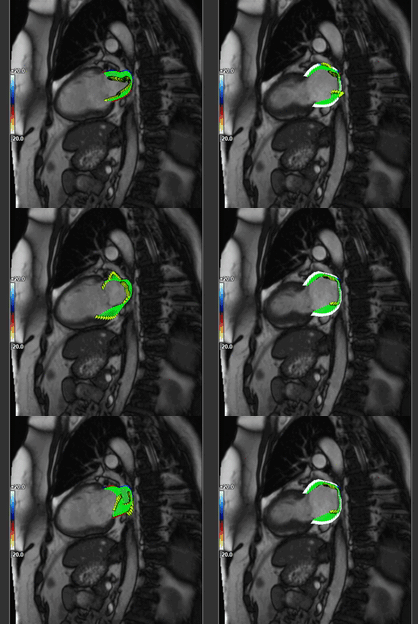
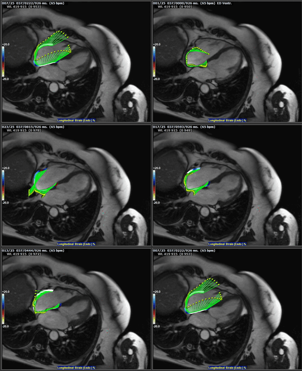
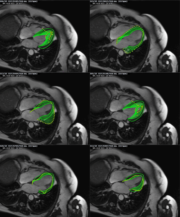
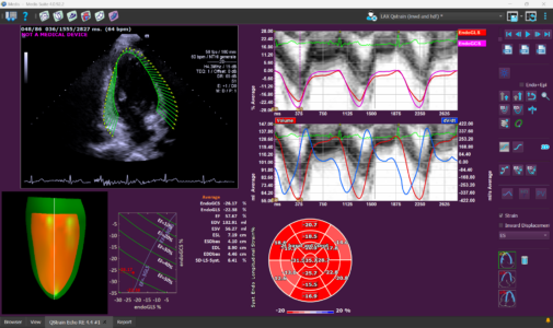
Understand myocardial deformation with Strain Analysis, a sensitive and reproducible way to quantify cardiac function from routine MR, CT and ultrasound images.
Ejection fraction tells you how much. Not how well.
For decades, cardiac function has been primarily assessed using volumetric measurements, especially ejection fraction (EF).
But cardiac dysfunction often starts before ejection fraction changes.
The myocardium continuously shortens, thickens and twists during every heartbeat. Measuring these subtle deformations provides insight into cardiac performance that cannot always be detected with volumetric measurements alone.
This is where myocardial strain comes in.
Strain measures myocardial deformation during contraction and relaxation, providing quantitative information about cardiac mechanics. Global Longitudinal Strain (GLS), Global Circumferential Strain (GCS) and Global Radial Strain (GRS) have become increasingly important markers for detecting early myocardial dysfunction, monitoring disease progression and evaluating treatment response.
Unlike traditional volumetric measurements, strain can reveal subtle abnormalities before ejection fraction declines, making it a valuable tool for diagnosis, treatment planning and patient follow-up.
Medis makes strain analysis accessible through a single, integrated workflow:
• Available for MR, CT and Ultrasound
• AI-powered contour detection*
• Vendor-independent tracking technology
• Automated strain calculation and reporting
• Global and regional analysis for ventricles and atria
Built on proven Speckle Tracking and Feature Tracking technology, Medis strain algorithms were developed by pioneers in the field of strain analysis. The vendor-independent algorithms have been optimized for each imaging modality and have been used in more than 1,700 scientific publications.
Note: Strain can be derived from data that is already acquired by default. In MR this is called CINE data. There is no need to acquire additional data for strain.
*AI automation is only available for MR images
One solution. Four chambers.
Most strain analysis focus primarily on the left ventricle. Medis goes further.
Analyze deformation across the left ventricle, right ventricle, left atrium and right atrium using a single, integrated workflow for MR, CT and ultrasound.
Medis QStrain provides a comprehensive view of cardiac mechanics (such as ventricular dysfunction, atrial remodeling etc.) that supports earlier detection, improved risk stratification and more informed clinical decision-making.
This approach allows clinicians to evaluate the heart as an interconnected system rather than a collection of individual chambers.
Clinical relevance
Strain analysis has demonstrated clinical value across a wide range of cardiovascular diseases:
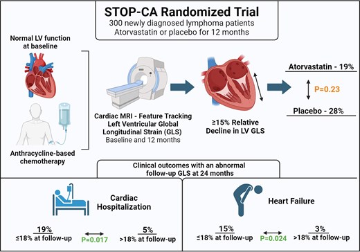
• Myocardial strain imaging enables early detection of anthracycline-related cardiac injury before substantial reductions in LVEF occur. In lymphoma patients receiving anthracycline chemotherapy, a follow-up GLS <18% predicted adverse cardiac outcomes, including heart failure and cardiac hospitalization.
• GLS assessment by cardiac magnetic resonance detected more CTRCD than EF criteria and showed greater sensitivity for predicting heart failure symptoms at 1 year. These findings suggest that GLS surveillance during chemotherapy may provide earlier detection of subclinical heart failure and cancer therapy-related cardiac dysfunction than EF alone.
STOP-CA Randomized Trial abstract summary (Juhasz et al.)
• CMR-derived LA reservoir strain is an independent predictor of new onset AF in HCM patients and provides incremental prognostic value beyond tradition risk factors. Incorporating LA strain into risk assessment may guide intensity of arrhythmia surveillance, and aid in early identification and treatment of high-risk patients.
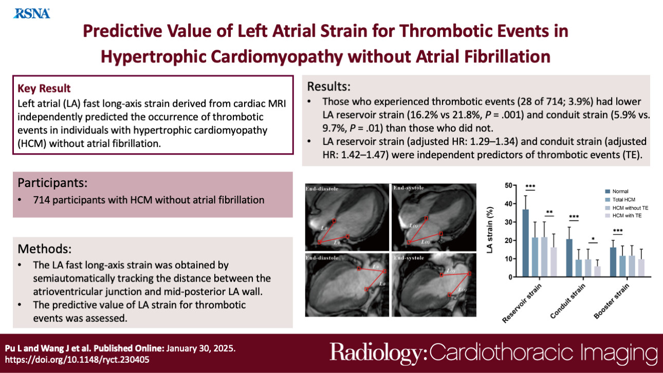
• Cardiac MRI–derived LA reservoir and conduit strain were independent predictors for the occurrence of TEs in individuals with HCM without AF.
• HCM patients with HFpEF exhibit both systolic and diastolic dysfunction, accompanied by increased diffuse and focal fibrosis. Independent predictors of HFpEF include lower LV-eGLSr, higher segmental ECV, atrial fibrillation, and drinking. The H2FPEF score shows significant associations with tissue-level abnormalities, highlighting the complementary role of CMR-derived strain and tissue characterization in the early detection and risk stratification of HFpEF in HCM.
• In STEMI patients with a concurrent CTO, strain significantly improves over time, regardless of CTO-PCI. Global strain is an independent predictor for functional recovery, incremental to infarct size, LVEF, and clinical parameters. Segmental strain was able to predict the recovery of wall thickening, incremental to transmural extent of infarction.
• LV global strains measured after primary PCI can predict the extent of myocardial recovery.
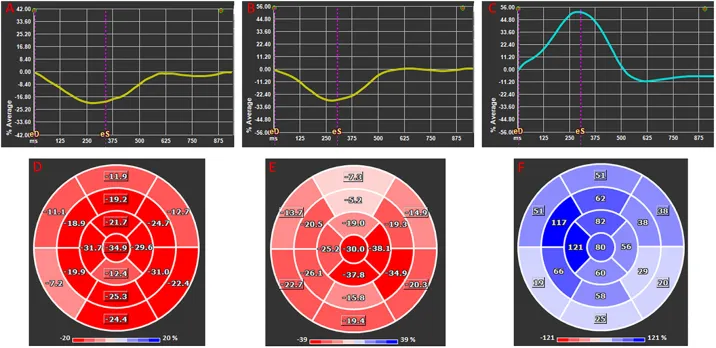
(Taha et al.) Global longitudinal (A,D), circumferential (B,E), and radial (C,F) 2D strain presented as average global strain curves (A–C) and corresponding segmental strain according to American Heart Association 17 segments Bull's eye ventricular maps (D–F). Note that longitudinal and circumferential strains are negative at their peak as they represent decrease in in fiber dimension whereas radial strain is positive as it infers increased myocardial thickness.
• Left ventricular global early diastolic longitudinal strain rate obtained from cardiovascular MRI feature tracking was independently associated with adverse outcomes in patients with heart failure with preserved ejection fraction.
• HCM patients with HFpEF exhibit both systolic and diastolic dysfunction, accompanied by increased diffuse and focal fibrosis. Independent predictors of HFpEF include lower LV-eGLSr, higher segmental ECV, atrial fibrillation, and drinking. The H2FPEF score shows significant associations with tissue-level abnormalities, highlighting the complementary role of CMR-derived strain and tissue characterization in the early detection and risk stratification of HFpEF in HCM.
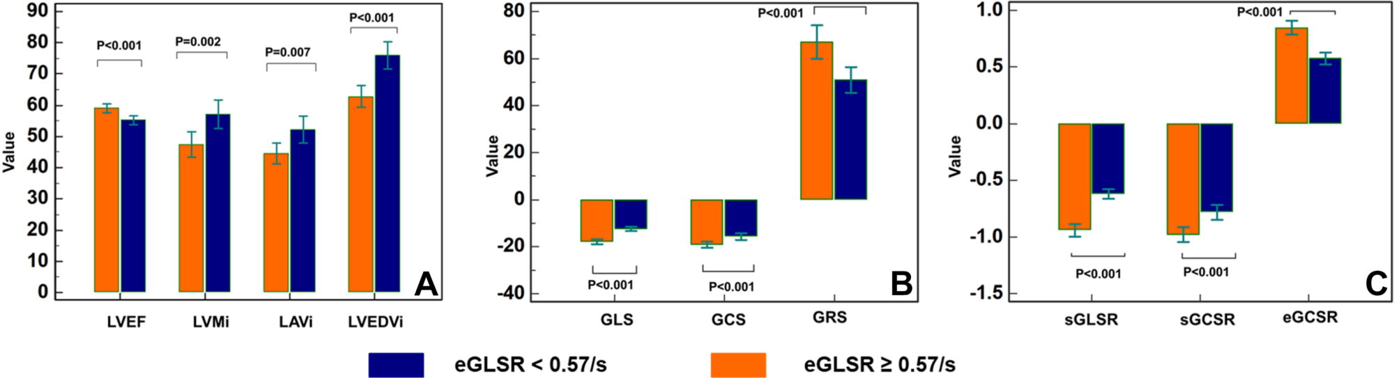
(He et al.) The patient cohort was divided into two groups according to the median global early diastolic longitudinal strain rate (eGLSR). (A) Structural and functional parameters, (B) strain parameters, and (C) strain rates showed significant difference between the two groups (the lower the eGLSR, the poorer the cardiac function). eGCSR = global early diastolic circumferential strain rate, GCS = global circumferential strain, GLS = global longitudinal strain, GRS = global radial strain, LAVi = left atrial maximum volume index, LVEDVi = left ventricular end-diastole volume index, LVMi = left ventricular end-diastole mass index, LVEF = left ventricular ejection fraction, sGCSR = global systolic circumferential strain rate, sGLSR = global systolic longitudinal strain rate.
One of the unique strengths of Medis QStrain is its availability across multiple imaging modalities.
Using the same biomechanical principles and tracking methodology, Medis enables strain assessment on:
• Cardiac MRI
• Cardiac CT
• Echocardiography
This allows clinicians to obtain consistent cardiac function insights regardless of the imaging modality available for a particular patient.
Cardiac MRI
Cardiac CT
Echocardiography
AI-powered workflow
Accelerate strain analysis with intelligent automation. AI-powered contouring automatically detects endocardial and epicardial borders, helping reduce analysis time while improving reproducibility. Automation is embedded throughout the workflow, including series detection and loading, view recognition, strain calculation, reporting, and export of results in JSON, XML, and DICOM SR formats. The result is a streamlined workflow that lets clinicians spend less time on image processing and more time on patient care.
Why clinicians choose Medis Strain?
Non-invasive
No catheterization or additional imaging
Workflow-friendly
Uses routine clinical images
Multi-modality
MR, CT and Ultrasound
Proven tracking technology
Algorithms used in over 1700 scientific publications
AI-powered
High reproducibility with minimal manual interaction
Trusted by leading physicians
“In our cardiac imaging research, strain analysis has proven to be a valuable tool for gaining insights into myocardial function beyond ejection fraction and volumetric assessment, across both ventricles and atria. Medis enables strain analysis in both cardiac MRI and echocardiography through intuitive and automated workflows, ensuring efficient and, most importantly, reproducible analysis. This streamlined approach highlights how advanced strain assessment can transition from a primarily research-based application into clinical practice, supporting broader adoption in routine patient evaluation and follow-up.”
Alexander Schulz, MD, FESC, FEACVI
Research Group Leader Multimodality Cardiovascular Imaging in Cardiology