User testimonials
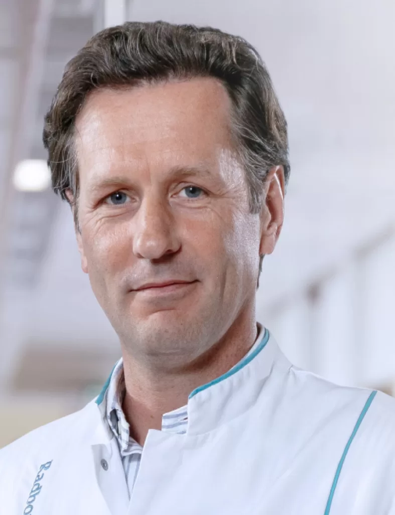
Professor of Cardiovascular Imaging and Cardiologist at Radboud University Medical Center, Nijmegen, the Netherlands.

Director of Cardiovascular Medicine Institute at Tec Salud, technologico de Monterrey Hospital Zambrano Hellion, Monterrey, Mexico
Erasmo de la Pena-Almaguer is a Cardiologist with postdoctoral training in Nuclear Cardiology, MAgnetic Resonance Imaging and Cardiovascular Comouted Tomography, with a fellowship both at Brigham and Womens Hospital, Harvard Medical School and Stanford University Medical Center Founding member of SCCT and Fellow of the American College of Cardiology as well ad the SCCT He currently is the director of the Cardiovascular Medicine Institute at Tec Salud, Tecnológico de Monterrey Hospital Zambrano Hellion in Monterrey Mexico
How long have you been using Medis Suite MR and what aspects attracted you to this product?
I have been using Medis post processing solutions for more than 15 years and what has attracted me is the ease of use and the great amount of information that it provides.
What are the benefits that you experience from using Medis Suite MR?
What I always saw as a big benefit is the super expedited processing, with all the tools needed for a comprehensive cardiac MR report.
What are your thoughts on the newly released novel parameter, Inward Displacement?
Inward Displacement is a great new tool that will allow to quantify wall motion abnormalities in an objective way. I recently published a case report demonstrating the use of Inward Displacement to objectively confirm regional dysfunction in a patient with a myocardial infarct.
What made Medis (Suite MR) stand out from other options in the market?
For me it is the ease of use and having a total comprehensive tool for every type of pathology analysed using CMR.
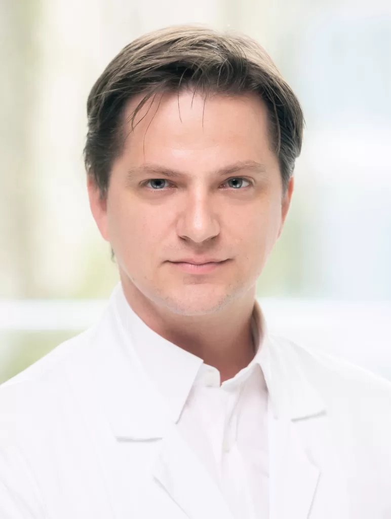
Dr. Dietrich Beitzke leads the cardiovascular MR Unit at the Department of Biomedical Imaging and Image-guided Therapy of the Medical University of Vienna. His research focuses on cardiac MR after PD. Dr. Dietrich Beitzke leads the cardiovascular MR Unit at the Department of Biomedical Imaging and Image-guided Therapy of the Medical University of Vienna. His research focuses on cardiac MRI after transplantation and hybrid PET/MR in cardiovascular diseases.
How long have you been using Medis Suite MR and what aspects attracted you to this product?
We are using Medis Suite MR since 2016 for our research and in clinical routine. The most important aspect is the improvement of our CMR post-processing workflow in terms of time and accuracy.
What are the benefits that you experience from using Medis Suite MR? How did it change your day-to-day practice?
Medis Suite MR really improved our clinical workflow especially with implementation of the robust automated ventricular contouring and the clearly arranged reporting function.
What are your thoughts on QMap within Medis Suite MR and in what way did QMap change/affect your daily workflow?
QMap improved especially the work up of newly diagnosed left ventricular hypertrophy and improved our capability to monitor treatment response in storage diseases like cardiac amyloidosis and Fabry Anderson Disease.
What made Medis (Suite MR) stand out from other options in the market?
We are very satisfied with the customer service Medis is providing. The after sales service is always easily accessible.
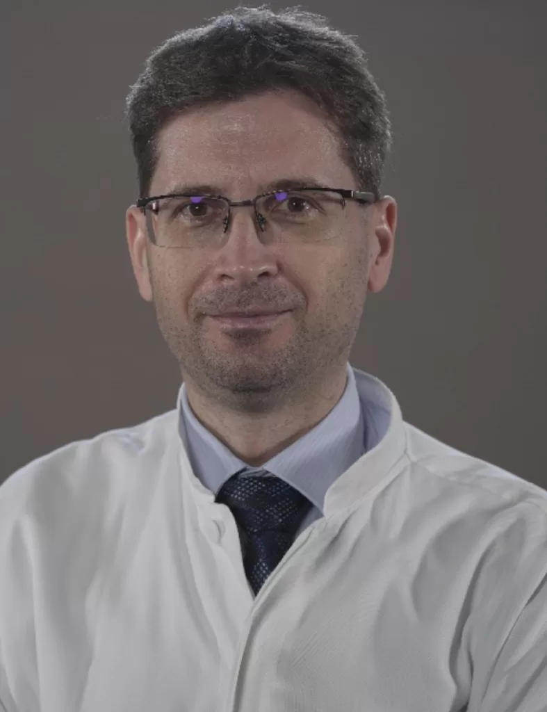
Professor Atilla Toth is a radiologist and cardiac MR specialist with 20 years of experience with interest areas of congenital heart disease, GUCH/ACHD. Staff member from 2004 at the Heart and Vascular Center of Semmelweis University. Radiology Board Certification in 2008. Level 3 EACVI certified CMR and CHD CMR specialist.
How long have you been using Medis Suite MR and what aspects attracted you to this product?
I’ve started my CMR career on a 1.5T GE scanner. The console was running IRIX on an Octane 2 and by that time Advantage Workstation 2.0 was running Solaris on Sparc hardware. Medis QMass 3.0 and QFlow were delivered as the evaluation software for cardiac use. I’ve been using Medis since than. I’ve used different versions of Medis on Linux and eventually on Windows. The graphical user experience is an important aspect for me, for example rubber-banding.
What are the benefits that you experience from using Medis Suite MR?
It is good to speed up the workflow using automated modules and options, but it is still important to be able to interact and modify a given step if I’d like to do so. If the workflow is too strict/hard-wired or it is hard to adjust the automatic end-result upon need, I prefer the alternative that lets me play.
What are your thoughts on the newly released novel parameter, Inward displacement?
QStrain is a quick and easy solution, but I’ve found the bull’s eye visualization not intuitive enough in a lot of cases.
On the other hand, Inward Displacement module with it’s simple mechanical concepts reflects wall motion abnormalities better in my opinion. It can be especially helpful for those who are less experienced or experts who have to supervise multiple cases.
What made Medis (Suite MR) stand out from other options in the market?
If I have to mention a useful feature, I’d emphasize AutoQ contour detection and mapping co-registration for easy ECV evaluation. I think Medis is easy to integrate, it runs on almost any hardware (true for both the server and the clients) and several colleagues can work concurrently with the floating license mechanism. Session files stores calipers and by a click of a button it brings me to the place of measurement in 3DView.
“We are excited to launch Medis QFR® in the VA Connecticut Cardiac Catheterization Laboratory. This innovative technology helps us provide cutting-edge, guided care to our Veterans with coronary artery disease.”
Interventional cardiologist / Cardiologist

This technique is simpler, safer and less expensive with equivalent outcomes and will conceivably be readily and widely adopted. Angiographically derived FFR, and specifically QFR®, should, in the future, be used routinely, reserving the invasive wire-based FFR approach for the challenging minority of cases where QFR® accuracy is questioned.
Interventional cardiologist / Cardiologist

“State-of-the-art approach in acute coronary syndrome targets on the “pancoronary risk”. This can be assessed easily, safely, and reproducibly by QFR®.”
Interventional cardiologist / Cardiologist

Interventional cardiologist / Cardiologist

“QFR® is a robust technology and it provides good diagnostic information and guiding information”
Clinical Researcher

“Medis Suite MR has increased our ease of use, saved us a lot of time, and consequently increased the quality of clinical reporting.”
Cardiologist

“Inward displacement is a great new tool that will allow to quantify wall motion abnormalities in an objective way”
Cardiologist

“Medis Suite MR is automated, but also gives the flexibility to interact”
Radiologist

“Medis Suite MR is our primary solution for reading CMR studies. Medis provides reliable and easy to use tools. We review cases with the team each day and find the viewer extremely valuable for these review sessions.”
Cardiologist

“Medis Suite MR improved our CMR post-processing workflow in terms of time and accuracy.”
Radiologist

“Medis Suite MR is our primary solution for reading CMR studies. Medis provides reliable and easy to use tools. We review cases with the team each day and find the viewer extremely valuable for these review sessions.”
Radiologist

“We have been studying coronary plaque using Medis Suite CT in patients with familial hypercholesteremia and were able to find that amount of plaque burden correlates with the estimated cardiovascular risk and number of future clinical events. This may be highly relevant for future development of personalized lipid lowering treatment. With help of the robust Medis Quantitative Plaque Analysis tools we were able to obtain our results in an efficient and highly reproducible manner.”
Director cardiovascular imaging

“Assessment of left ventricular global longitudinal strain on CT data with Medis Suite CT is very fast and reproducible, providing an additional piece of information in the risk stratification of patients undergoing to TAVI.”
Head of department of cardiovascular imaging

“We have been using the Medis Suite XA in our core laboratory since 1991. All major interventional device trials with angiographic endpoints have been evaluated using Medis Suite XA as the standard.”

“Medis Suite XA has proven to be very intuitive and easy to learn, even with the challenging cases in our large real-world population trial. Medis Suite XA allows us to perform a high volume of quantitative angiographic analyses with limited personnel.”

“Our technology offerings will allow Medis to complement and further enhance its already impressive suite of clinical offerings and spur innovation.”
Former CEO of Amid
“QIvus RE’s semi-automatic contour detection is excellent making it easy to quickly calculate volumetric and VH data.”
Manager Intravascular Imaging Laboratory, Cardiovascular Research Foundation

“Current QIvus RE software greatly shortened the time taken for OCT analysis. This is a big progress for us.”
Japan