Regional left ventricular wall motion can be assessed in different ways: by visual assessment, regional strain, and the new Medis innovative parameter Inward Displacement. All methodologies aim to identify regional myocardial impairment before it becomes manifest.
More specifically, although ejection fraction and global strain have shown to be of great value for clinical diagnosis, assessing regional dysfunction remains a challenge, even with regional strain. To assess regional dysfunction in a more objective and quantitative manner, we have introduced “Inward Displacement”.
Early feedback confirms the method works very well in MR, CT as well as in Echocardiography. With the Medis Medical Imaging Solution, you can quickly and accurately quantify the left ventricular regional wall motion.
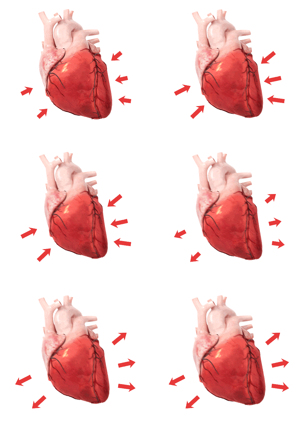
Regional strain provides information on regional ventricular dysfunction by measuring the changes in parallel displacement along the left ventricular circumference. However, the sensitivity of regional strain has been shown to be relatively low. This is due to the fact, that small changes in the length of a subsegment along the perimeter of the left heart chamber are prone to large variabilities, depending on the definitions of these subsegments.
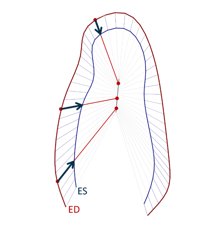
Over the years, various left ventricular wall motion models have been developed, the most well-known model is the centerline model [1]. This model is based on the changes in position of individual points along the LV perimeter from ED to ES and perpendicular to the centerline between the ED and ES contours. However, this is method not based on a solid wall motion model.
The feature tracking technology provides much more information about the actual displacement of the individual positions along the left ventricular boundary. Based on that solid technology, Medis has created an actual left ventricular wall motion model which shows that points along the endocardial border are moving towards well-defined “centers of contraction”. The new Inward Displacement model allows for an objective quantitative assessment of regional function. Figure 1 shows a graphical representation of the basic principles of the Inward Displacement model.
Over the years, strain has demonstrated its value for the function of the left ventricle in different cardiac disorders such as myocardial infarction, cardiac toxicity, and various cardiomyopathies. Some of the pathologies are highlighted below.
The value of strain keeps increasing by new evidence that is gathered through research highlighting the incremental prognostic value of strain in various cardiac disorders
To detect myocardial infarction, it is expected that changes in the regional strain and inward displacement are visible in multiple segments, in line with the associated coronary trajectories.
For example, the CT-derived regional strain was significantly reduced in patients with significant disease in the LAD compared with healthy controls [2]. Also, with MR [3] and Ultrasound [4], the ability to detect myocardial infarcts based on regional wall motion technologies has been illustrated.
Recently, a case report was published demonstrating the use of Inward Displacement to objectively confirm regional dysfunction in a patient with a myocardial infarct [5]. Assessing left ventricular remodeling and defining therapeutic strategies could be done based on the regional wall motion results.
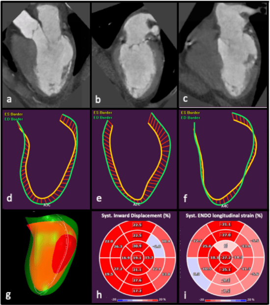
Due to regional strain, cardiac amyloidosis can be diagnosed by the presence of relative apical sparing [6]. Regional strain by echocardiography can be measured quantitatively in which the apical regional strain shows normal values. This can also be seen in figure 2 in which normal apical regional strain values can be seen whereas basal and mid-regional strain values are reduced. Another article shows that regional strain quantification enables differentiating cardiac amyloidosis from other pathologies such as hypertrophic cardiomyopathy [7].

There has been a great need to describe accurately and quantitatively regional left ventricular wall motion along its actual motion trajectories, and not along the perimeter of the chamber, which does not describe the actual physiology. For that reason, Medis has, together with our KOLs, developed the Inward Displacement model as a new innovative methodology based on Speckle or Feature tracking, as well as a model that describes better the actual trajectories rather than the centerline method. Therefore, the Inward Displacement bull’s eye plots describe the regional impairment most in line with visual observations.
Our Medis Inward Displacement solution is feasible for the Left Ventricle and is available for MR, CT, and Ultrasound to further enhance insights for your patients’ cases.
Inward Displacement across modalities
Understanding Ultrasound: Strain
Medis’ tracking algorithms are created by the Founders of strain. Both the speckle and feature tracking algorithms are robust, vendor-independent, and proven algorithms. The algorithms have been optimized for each specific modality. Altogether, our tracking algorithms have been used in over 1700 scientific papers.
The inward displacement measurement is based on Feature Tracking hence we have incorporated it in the Strain application. Once you have performed your strain analysis, you are one button click away from your Inward Displacement results presented in the Bull’s Eye plots. The values will be presented based on your regular cine images without any additional time or effort.
Inward Displacement can be measured on Ultrasound, MRI, and CT images with Medis. This enables comparing data from one modality to another, as for all modalities the inward displacement is based on the same algorithm., Medis multimodality solution, ensures that very likely you will have inward displacement data for each patient, depending on at least one available modality
Getting the long axis endocardial borders can be a time-consuming and repetitive task. For MR data deep learning contour detection has been embedded, for which we have had great feedback from our users. The Medis AI Deep learning algorithm, which has been trained on almost 900 datasets in various cases across different vendors and pathologies, increases reproducibility.
Inward Displacement can be measured in Ultrasound, MR and CT images. Medis multimodality solution ensures that very likely you will have inward displacement data for each patient, depending on at least one available modality
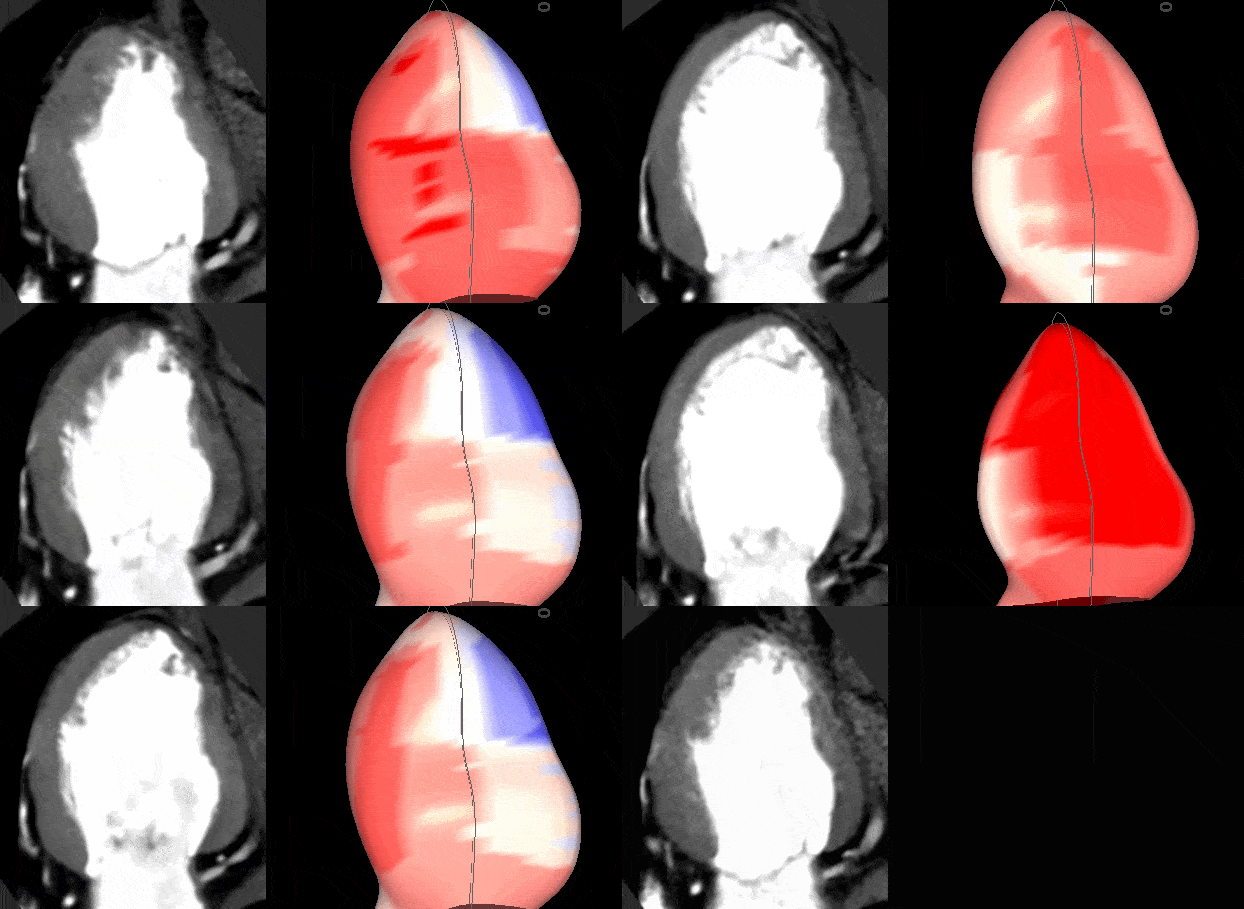
Medis Suite CT
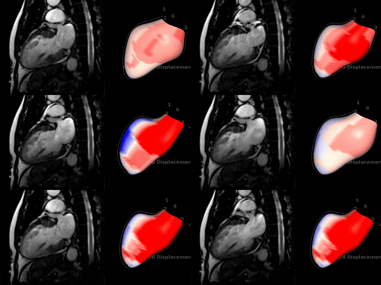
Medis Suite MR
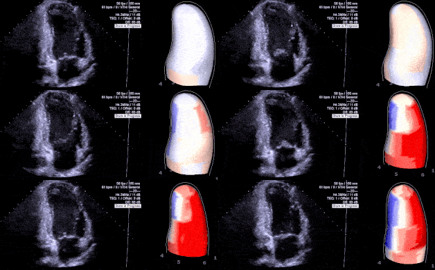
Medis Suite UltraSound

"The newly released novel parameter, Inward Displacement is a great new tool that will allow quantifying wall motion abnormalities in an objective way."
Prof. Erasmo de la Peña Director of the cardiovascular Medicine Institute at Tec Salud, Tecnológico de Monterrey Hospital Zambrano Hellion in Monterey, MexicoDo you want to learn more about Medis products? Contact us and we can set up a demo.
"QStrain is a quick and easy solution, but I've found the bull's eye visualization not intuitive enough in a lot of cases. On the other hand Inward Displacement module with it's simple mechanical concepts reflects wall motion abnormalities better in my opinion. It can be especially helpful for those who are less experienced or experts who have to supervise multiple case."
Dr. Atilla Tóth Radiologist at Gottsegen György Hungarian Institute of Cardiology & Semmelweis University