Highly reliable and robust quantitative analysis – the nr.1 vendor-independent choice of many departments for more than 15 years. The ventricular function module has supported clinical research in over 650+ peer reviewed publications
Deep learning algorithm for fully automatic Left Ventricle and Right Ventricle contours
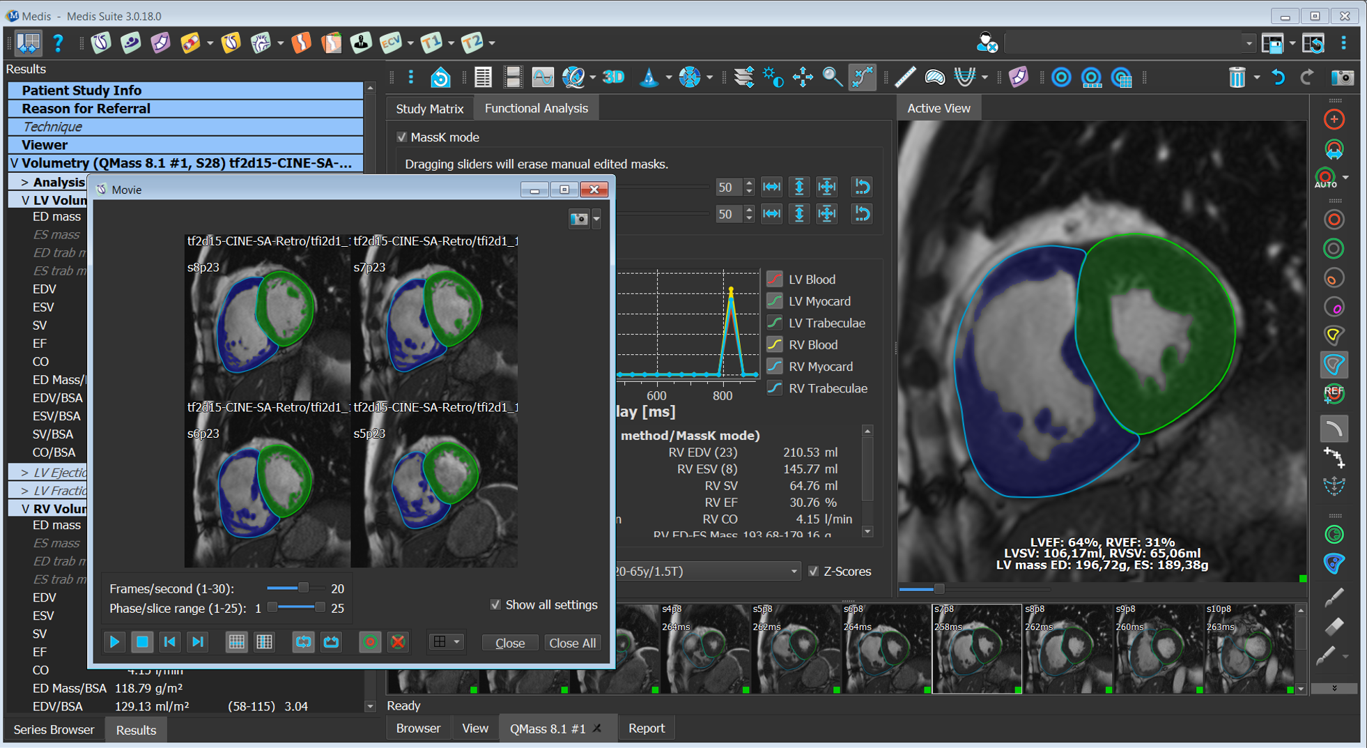
The user interface makes function analysis easy and practical
‘LiveContour’ algorithm for quick semi-automatic LV contours
‘MassK’ algorithm which allows for quick exclusion of papillary muscle and trabeculations of the left and right ventricle
Function is also possible with CT using the exact same post processing method. Because of the streamlined analysis workflow, Medis Suite allows you to compare CT and MR function studies with the highest diagnostic confidence
Contours traced during the left ventricle function analysis can be loaded into the strain application for subsequent deformation analysis based on features tracking
Features
Global Function Analysis
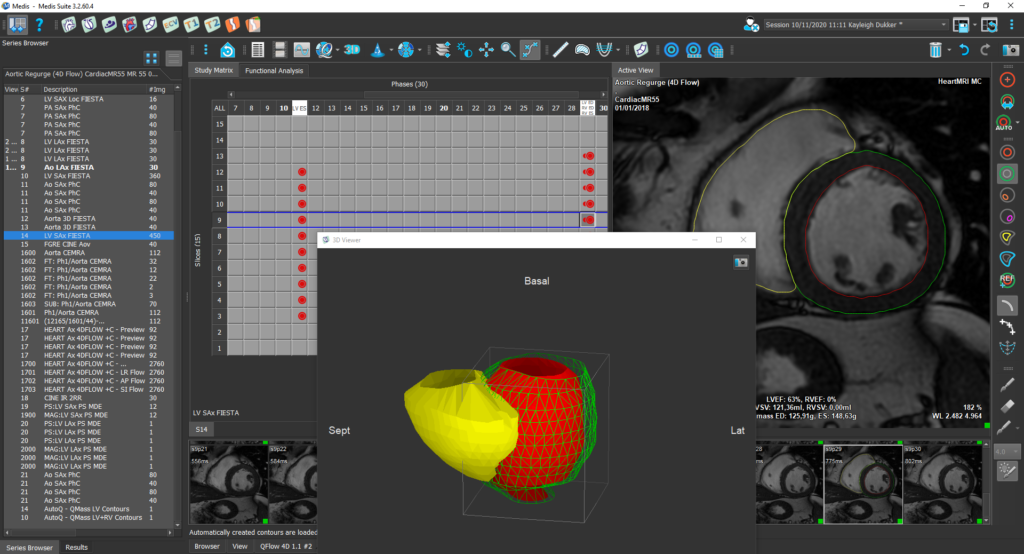
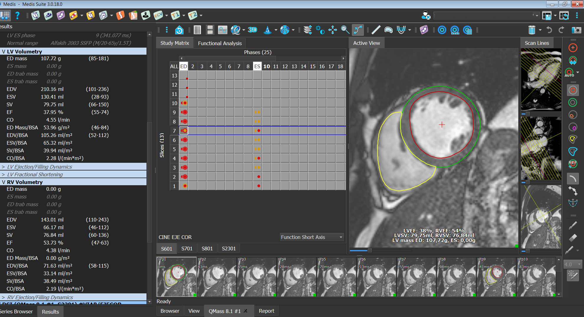
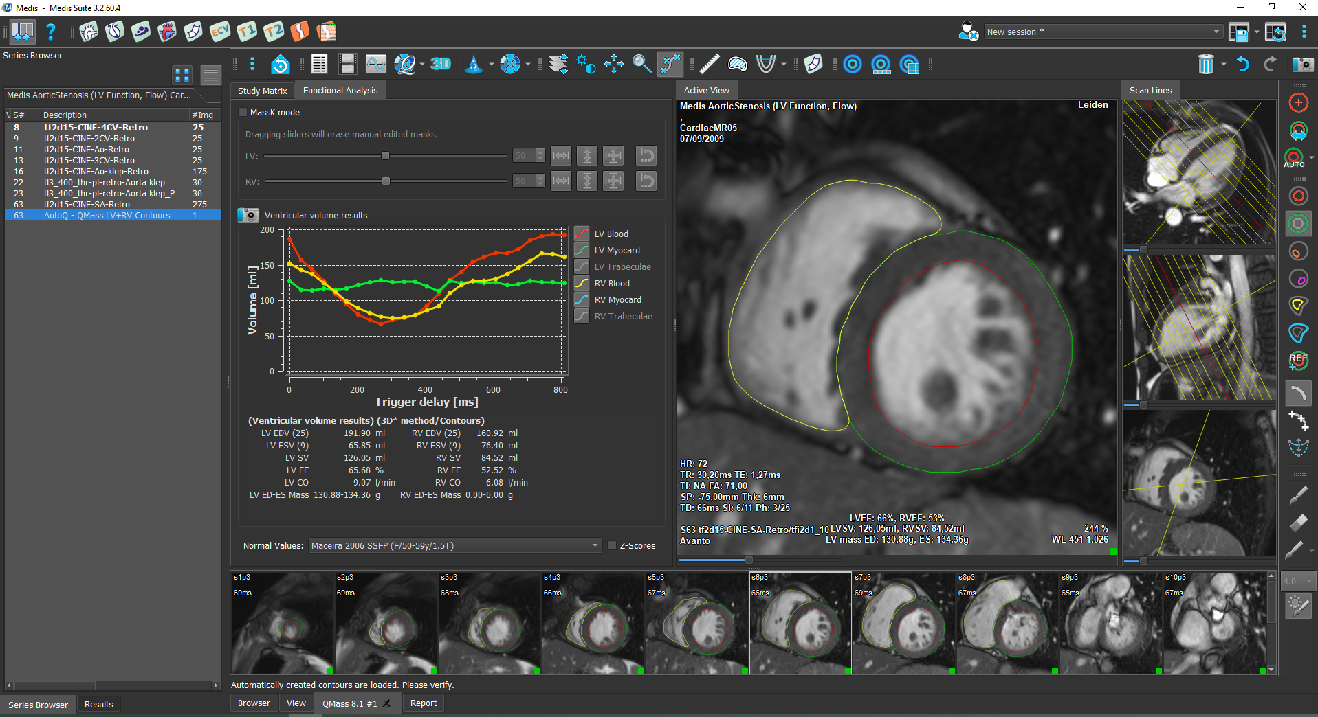
Left ventricle (LV) and right ventricle (RV) volume analysis to get parameters as ejection fraction and stroke volume
AI based automatic LV & RV contour detection algorithm to minimize manual corrections for short and long axis images
Enhanced guided workflow for a quick analysis
Quantification of custom volumes, such as atrial volumes
Absolute and BSA indexed results to normalize the results
Provides normal ranges based on academic papers to compare results with the same age and gender
Provides z-scores for pediatric cases in the results
Area-length and Biplane analysis method for long axis function analysis
‘MassK’ algorithm available for automatic segmentation of papillary muscles and trabeculations
Regional Function Analysis
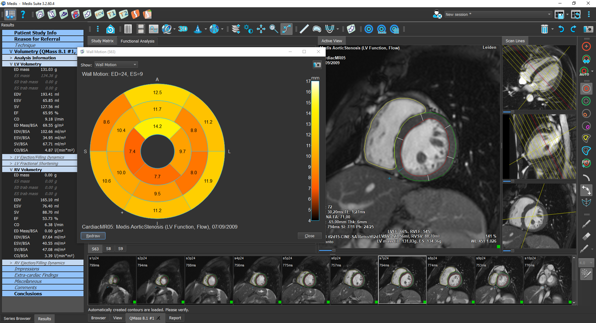
Wall motion analysis to analyse the uniformity
Wall thickness and wall thickening measurements
Wall thickness changes over time to analyse the contractility
Results are displayed within curves or AHA 16 segment bull’s eye
Other features
Visual scoring for wall motion, perfusion, and infarct size in the 16 segments of the heart
Benefits
Highly reliable and robust quantitative analysis – the nr.1 vendor-independent choice of many departments for more than 15 years. The ventricular function module has supported clinical research in over 650+ peer reviewed publications
Deep learning algorithm for fully automatic Left Ventricle and Right Ventricle contours
The user interface makes function analysis easy and practical
‘LiveContour’ algorithm for quick semi-automatic LV contours
‘MassK’ algorithm which allows for quick exclusion of papillary muscle and trabeculations of the left and right ventricle
Function is also possible with CT using the exact same post processing method. Because of the streamlined analysis workflow, Medis Suite allows you to compare CT and MR function studies with the highest diagnostic confidence
Contours traced during the left ventricle function analysis can be loaded into the strain application for subsequent deformation analysis based on features tracking
Features
Global Function Analysis
Left ventricle (LV) and right ventricle (RV) volume analysis to get parameters as ejection fraction and stroke volume
AI based automatic LV & RV contour detection algorithm to minimize manual corrections for short and long axis images
Enhanced guided workflow for a quick analysis
Quantification of custom volumes, such as atrial volumes
Absolute and BSA indexed results to normalize the results
Provides normal ranges based on academic papers to compare results with the same age and gender
Provides z-scores for pediatric cases in the results
Area-length and Biplane analysis method for long axis function analysis
‘MassK’ algorithm available for automatic segmentation of papillary muscles and trabeculations
Regional Function Analysis
Wall motion analysis to analyse the uniformity
Wall thickness and wall thickening measurements
Wall thickness changes over time to analyse the contractility
Results are displayed within curves or AHA 16 segment bull’s eye
Other features
Visual scoring for wall motion, perfusion, and infarct size in the 16 segments of the heart
LV/RV function in use
Book a demo of Medis Suite MR
You are only a few clicks away from having a demo with an experienced Medis Suite MR product specialist