Unlock CT-derived coronary plaque insights for informed interventions.
A highly automated analysis performed on any existing
coronary CTA.
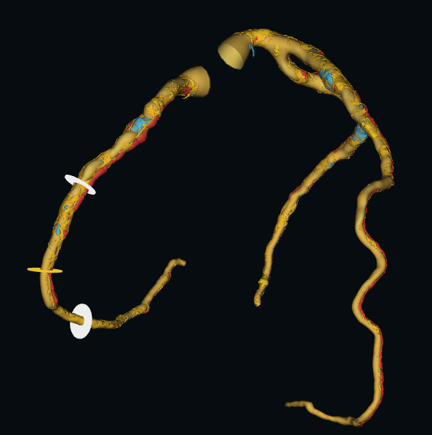
QGuideCT1 extracts and labels the coronary tree automatically, including main epicardial vessels and side
branches, and renders a 3D reconstructed visualization of the analyzed vessels which can be made actionable for determining treatment paths.
1QGuideCT is a registered trademark of Medis Associated BV.
QGuideCT 1.0 has received US market clearance from the FDA.
CE certification pending for this device. Not for clinical use in the EU.
QUESTION 1: Where is the plaque and what is it made of?
• The whole tree, not one slice. Plaque is quantified continuously along the vessel rather than judged from a single cross-section. • Automated extraction of the coronary tree and centreline, with automated vessel labelling. • Reader stays in control. Intuitive and easy to use longitudinal and transversal contour editing possible when needed.
Plaque composition colour key
Low density lipid-rich morphology
Non-calcified soft / fibrous burden
Calcified drives interventional strategy
Reported per vessel: volume by component (mm³) · total plaque (mm³) · lesion length (mm) · plaque burden (%) plus a total plaque volume across all vessels of interest.
3D reconstructed visualization from a standard coronary which enables intuitive visualization of the plaque distribution as demonstrated in the P4 trial.
QUESTION 2: How does that change the treatment plan?
• Plan the case, not just the diagnosis. The 3D reconstruction aids in determining treatment paths before the patient is on the table.
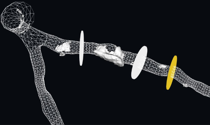
• Calcium strategy decided in advance. Where the calcium sits along the lesion informs whether lesion preparation is needed, and which kind. • Sizing and landing zones. Lesion length and plaque burden are measured on CT rather than estimated during the procedure.
What reaches the table
• Intuitive 3D Visualization
• Plaque burden
• CAD-RADS 2.0 report
• Wireframe view with calcium distribution
• Patient-specific angiographic views
• Lesion length measurements
Cath Lab View: the wireframe view easily shows the extend and distribution of calcium along the vessels.