Understand the forces driving cardiac function with Hemodynamic Forces, a non-invasive way to quantify intraventricular pressure gradients from routine MR, CT and ultrasound images.
*Hemodynamic Forces is for research use only and cannot be used in daily clinical practice for patient diagnosis and treatment.
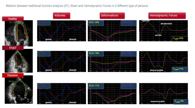
Ejection fraction tells you how much. Not how.
For decades, cardiac function has been defined by volumetrics, primarily ejection fraction.
But cardiac function is more than volume alone.
Blood flow is driven by pressure gradients, shaped by myocardial motion, and influenced by ventricular mechanics.
To truly understand cardiac function, you need to measure Hemodynamic Forces.
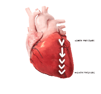
Each heartbeat creates pressure differences inside the ventricle that drive blood flow. These are known as intraventricular pressure gradients.
Hemodynamic Forces represent the global effect of these gradients across the entire ventricle.
Hemodynamic Forces add a new dimension to cardiac assessment:
• Efficiency of ventricular filling and ejection
• Direction and magnitude of forces driving blood flow
• Interaction between myocardial motion and intracardiac flow
This adds a functional layer to cardiac assessment, complementing volumetrics and strain.
Derived directly from routine MR, CT or ultrasound images, no additional scans required.
Simple workflow:
Acquire standard MR, CT or ultrasound cine images
Automatic AI-based contouring*
Endocardial motion tracking
Biomechanical modeling
Results visualized as force vectors and quantitative metrics
Medis makes Hemodynamic Forces accessible in daily practice:
• No additional scans
• No contrast agents
• Based on standard long-axis views
• Integrated into existing workflows
Clinical relevance
Hemodynamic Forces have shown promising value across multiple clinical scenarios:
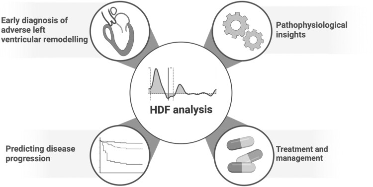
• LV HDF may significantly improve the detection of early myocardial systolic dysfunction when volumetric and deformation cardiac measures are still intact.
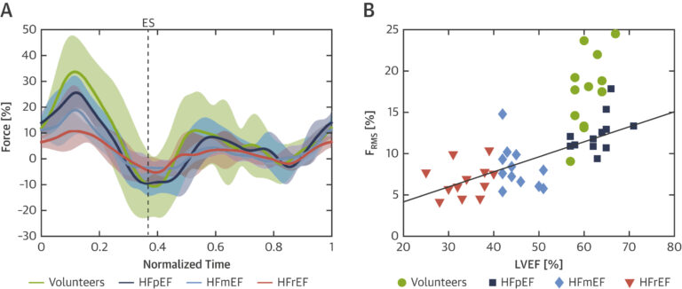
• Assessment of HDF indicates impairment of LV systolic ejection force in HFpEF, which is associated with cardiovascular events
• Misalignment of diastolic HDF after STEMI is associated with adverse LV remodeling after 4 months
• CMR-derived LV IVPG are univariably associated with MACE. However, LV IVPGs do not add prognostic value to LV ejection fraction and LV GLS
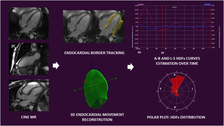
Filomena et al., From left to right: Cine CMR long axis data sets are used for left ventricular haemodynamic forces estimation. End-systolic and end-diastolic borders are traced and tracked frame-by-frame to allow endocardial border movement reconstruction in a three-dimensional model. Apex-to-base and latero-septal haemodynamic forces are estimated over time and graphically represented as curves. Haemodynamic forces distribution in a selected period of time is represented using a polar plot. 2C, two chamber view; 3C, three chamber view; 3D, three-dimensional; 4C, four chamber view; A, apex; B, base; ED, end-diastole; ES, end-systole; HDFs, haemodynamic forces; L, lateral wall; MR, magnetic resonance; S, septum.
• Dilated Cardiomyopathies: in the absence of pressure reversal, lower systolic ejection force, E-wave decelerative force, and overall LV IVPG are powerful predictors of outcome, independent of clinical and imaging parameters
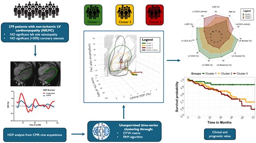
• Analyzing both longitudinal and transversal HDF throughout the cardiac cycle enables the identification of distinct phenotypes with prognostic value beyond EF and LGE in non-ischemic LV cardiomyopathy
Eur Heart J Cardiovasc Imaging, Volume 26, Issue 4, April 2025, Pages 630–639 (Airale et al.)
• Despite no further significant decline in LVEF and GLS after 3 months of anthracycline therapy, HDF continueto decrease, reflecting subtle changes in LV preceding overt functional impairment
• Higher sensitivity of HDFs as biomarkers of cardiotoxicity of cancer-related therapy
*Hemodynamic Forces is for research use only and cannot be used in daily clinical practice for patient diagnosis and treatment.
Hemodynamic Forces Example
Why clinicians choose Medis Hemodynamic Forces?
Non-invasive
No catheterization or additional imaging
Workflow-friendly
Uses routine clinical images
Multi-modality
MR, CT and Ultrasound
Proven tracking technology
Algorithms used in over 1700 scientific publications
AI-powered
High reproducibility with minimal manual interaction
Trusted by leading physicians
“We aim through research to better understand the underlying disease causes which can improve diagnosis or treatment in the end. With the HDF module of Medis, we have looked at patients with precapillary pulmonary hypertension to see if this parameter can help in the detection of subtle changes in the LV mechanics, which seems to be the case. This result makes us continue to further research the value of HDF.”